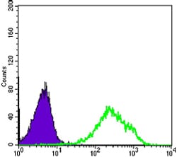
DAXX Primary Antibody

Item Information
Catalog #
Size
Price
Description

DAXX (death-domain associated protein), it is a multifunctional protein that resides in multiple locations in the nucleus and in the cytoplasm. It interacts with a wide variety of proteins, such as apoptosis antigen Fas, centromere protein C, and transcription factor erythroblastosis virus E26 oncogene homolog 1. In the nucleus, the encoded protein functions as a potent transcription repressor that binds to sumoylated transcription factors. Its repression can be relieved by the sequestration of this protein into promyelocytic leukemia nuclear bodies or nucleoli. This protein also associates with centromeres in G2 phase. In the cytoplasm, the encoded protein may function to regulate apoptosis. The subcellular localization and function of this protein are modulated by post-translational modifications, including sumoylation, phosphorylation and polyubiquitination. Alternative splicing results in multiple transcript variants.

Product Overview
Entrez GenelD
1616
Aliases
DAP6; EAP1; BING2
Clone#
7A11
Host / Isotype
Mouse / IgG1
Species Reactivity
Human
Immunogen
Purified recombinant fragment of human DAXX expressed in E. Coli.
Formulation
Ascitic fluid containing 0.03% sodium azide.
Storage
4°C; -20°C for long term storage
Product Applications
WB (Western Blot)
1/500 - 1/2000
ICC (Immunocytochemistry)
1/200 - 1/1000
FCM (Flow Cytometry)
1/200 - 1/400
ELISA
1/10000
References
1. Cell. 1997 Jun 27;89(7):1067-76.
2. Biochem Biophys Res Commun. 2000 Dec 9;279(1):6-10.
3. Proc Natl Acad Sci U S A. 2004 Aug 17;101(33):12130-5.
Product Image
Western Blot
Figure 1: Western blot analysis using DAXX mouse mAb against K562 cell lysate (1).
Immunofluorescence analysis
Figure 2: Confocal Immunofluorescence analysis of PANC-1 cells using DAXX mouse mAb (green). Blue: DRAQ5 fluorescent DNA dye.
Immunofluorescence analysis
Figure 3: Confocal Immunofluorescence analysis of Hela cells using DAXX mouse mAb (green). Red: Actin filaments have been labeled with Alexa Fluor-555 phalloidin. Blue: DRAQ5 fluorescent DNA dye.
Flow cytometric
Figure 4: Flow cytometric analysis of Hela cells using DAXX mouse mAb (green) and negative control (purple).
For Research Use Only. Not for use in diagnostic procedures.