

DAPP1 Primary Antibody
Item Information
Catalog #
Size
Price
Description
DAPP1 (Dual Adaptor Of Phosphotyrosine And 3-Phosphoinositides) is a Protein Coding gene. Among its related pathways are Immune System and Downstream signaling events of B Cell Receptor (BCR). GO annotations related to this gene include phospholipid binding and phosphatidylinositol-3,4-bisphosphate binding.
Product Overview
Entrez GenelD
27071
Aliases
BAM32
Clone#
2F7A9
Host / Isotype
Mouse / IgG1
Species Reactivity
Human
Immunogen
Purified recombinant fragment of human DAPP1 (AA: 127-276) expressed in E. Coli.
Formulation
Purified antibody in PBS with 0.05% sodium azide
Storage
Store at 4°C short term. Aliquot and store at -20°C long term. Avoid freeze/thaw cycles.
Product Applications
WB (Western Blot)
1/500 - 1/2000
FCM (Flow Cytometry)
1/200 - 1/400
ELISA
1/10000
References
1.J Immunol. 2011 Oct 15;187(8):3972-8.
2.J Immunol. 2004 Nov 1;173(9):5601-9.
2.J Immunol. 2004 Nov 1;173(9):5601-9.
Product Image
Elisa
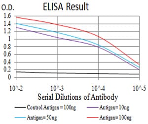
Figure 1: Black line: Control Antigen (100 ng);Purple line: Antigen (10ng); Blue line: Antigen (50 ng); Red line:Antigen (100 ng)
Western Blot
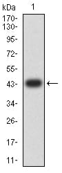
Figure 2:Western blot analysis using DAPP1 mAb against human DAPP1 (AA: 127-276) recombinant protein. (Expected MW is 43.5 kDa)
Western Blot
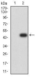
Figure 3:Western blot analysis using DAPP1 mAb against HEK293 (1) and DAPP1 (AA: 127-276)-hIgGFc transfected HEK293 (2) cell lysate.
Flow cytometric
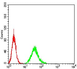
Figure 4:Flow cytometric analysis of A549 cells using DAPP1 mouse mAb (green) and negative control (red).
For Research Use Only. Not for use in diagnostic procedures.