

DAPK3 Primary Antibody
Item Information
Catalog #
Size
Price
Description
Death-associated protein kinase 3 (DAPK3) induces morphological changes in apoptosis when overexpressed in mammalian cells. These results suggest that DAPK3 may play a role in the induction of apoptosis.
Product Overview
Entrez GenelD
1613
Aliases
ZIP; ZIPK
Clone#
2H1D11
Host / Isotype
Mouse / IgG1
Species Reactivity
Human
Immunogen
Purified recombinant fragment of human DAPK3 (AA: 28-161) expressed in E. Coli.
Formulation
Purified antibody in PBS with 0.05% sodium azide.
Storage
Store at 4°C short term. Aliquot and store at -20°C long term. Avoid freeze/thaw cycles.
Product Applications
WB (Western Blot)
1/500 - 1/2000
IHC_P(Immunohistochemistry)
1/200 - 1/1000
FCM (Flow Cytometry)
1/200 - 1/400
ELISA
1/10000
References
1. Cancer Res. 2011 Apr 15;71(8):3152-61.
2. Int J Cancer. 2009 Apr 1;124(7):1587-93.
2. Int J Cancer. 2009 Apr 1;124(7):1587-93.
Product Image
Western Blot
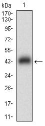
Figure 1: Western blot analysis using DAPK3 mAb against human DAPK3 (AA: 28-161) recombinant protein. (Expected MW is 41.6 kDa)
Western Blot

Figure 2: Western blot analysis using DAPK3 mAb against HEK293 (1) and DAPK3 (AA: 28-161)-hIgGFc transfected HEK293 (2) cell lysate.
Flow cytometric

Figure 3: Flow cytometric analysis of A431 cells using DAPK3 mouse mAb (green) and negative control (red).
Immunohistochemical analysis

Figure 4: Immunohistochemical analysis of paraffin-embedded rectum cancer tissues using DAPK3 mouse mAb with DAB staining.
Elisa

Black line: Control Antigen (100 ng); Purple line: Antigen(10ng); Blue line: Antigen (50 ng); Red line: Antigen (100 ng);
For Research Use Only. Not for use in diagnostic procedures.