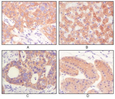
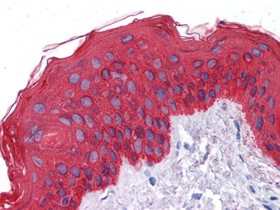
Cytokeratin (Pan) Primary Antibody

Item Information
Catalog #
Size
Price
Description

Biochemically, most members of the CK family fall into one of two classes, type I (acidic polypeptides) and type II (basic polypeptides). The type II cytokeratins consist of basic or neutral proteins which are arranged in pairs of heterotypic keratin chains coexpressed during differentiation of simple and stratified epithelial tissues. Cytokeratins comprise a diverse group of intermediate filament proteins (IFPs) that are expressed as pairs in both keratinized and non-keratinized epithelial tissue. Cytokeratins play a critical role in differentiation and tissue specialization and function to maintain the overall structural integrity of epithelial cells. Cytokeratins have been found to be useful markers of tissue differentiation which is directly applicable to the characterization of malignant tumors.

Product Overview
Entrez GenelD
3852
Aliases
K5; DDD; EBS2; KRT5A; KRT5
Clone#
7H8C4
Host / Isotype
Mouse / IgG1
Species Reactivity
Human
Immunogen
Purified recombinant fragment of CK5 expressed in E. Coli.
Formulation
Ascitic fluid containing 0.03% sodium azide.
Storage
4°C; -20°C for long term storage
Product Applications
WB (Western Blot)
1/500 - 1/2000
IHC_P(Immunohistochemistry)
1/200 - 1/1000
ICC (Immunocytochemistry)
1/200 - 1/1000
ELISA
1/10000
References
1. Scope A. Schwendenwein I. Frommlet F. Vet Rec. 2006, Dec 16, 159(25): 839-43.
2. Somjen D. Katzburg S. Posner GH. et al. J Cell Biochem. 2007, Apr 15, 100(6): 1406-14.
Product Image
Western Blot
Figure 1: Western blot analysis using CK mouse mAb against truncated CK5 recombinant protein.
Immunohistochemical analysis
Figure 2: Immunohistochemical analysis of paraffin-embedded human lung squamous cell carcinoma (A),normal hepatocyte (B), colon adenocacinoma?, normal stomach tissue (D), showing cytoplasmic and membrane localization using CK mouse mAb with DAB staining.
Immunohistochemical analysis
Figure 3: Immunohistochemical analysis of paraffin-embedded human Skin tissues using CK mouse mAb
Immunofluorescence analysis
Figure 4: Immunofluorescence staining of methanol-fixed Eca-109 (left) and HepG2 (right) cells showing cytoplasmic localization.
For Research Use Only. Not for use in diagnostic procedures.