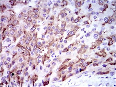
CYP3A4 Primary Antibody

Item Information
Catalog #
Size
Price
Description

This gene encodes a member of the cytochrome P450 superfamily of enzymes. The cytochrome P450 proteins are monooxygenases that catalyze many reactions involved in drug metabolism and synthesis of cholesterol, steroids and other lipids. This protein localizes to the endoplasmic reticulum and its expression is induced by glucocorticoids and some pharmacological agents. This enzyme is involved in the metabolism of approximately half the drugs in use today, including acetaminophen, codeine, cyclosporin A, diazepam and erythromycin. The enzyme also metabolizes some steroids and carcinogens. This gene is part of a cluster of cytochrome P450 genes on chromosome 7q21.1. Previously another CYP3A gene, CYP3A3, was thought to exist; however, it is now thought that this sequence represents a transcript variant of CYP3A4. Alternatively spliced transcript variants encoding different isoforms have been identified.

Product Overview
Entrez GenelD
1576
Aliases
HLP; CP33; CP34; CYP3A; NF-25; CYP3A3; P450C3; CYPIIIA3; CYPIIIA4; P450PCN1
Clone#
3H8
Host / Isotype
Mouse / IgG1
Species Reactivity
Human
Immunogen
Purified recombinant fragment of human CYP3A4 expressed in E. Coli.
Formulation
Purified antibody in PBS with 0.05% sodium azide
Storage
4°C; -20°C for long term storage
Product Applications
WB (Western Blot)
1/500 - 1/2000
IHC_P(Immunohistochemistry)
1/200 - 1/1000
ICC (Immunocytochemistry)
1/200 - 1/1000
FCM (Flow Cytometry)
1/200 - 1/400
ELISA
1/10000
References
Drug Metab Dispos. 2009 Dec;37(12):2305-13.
Biochem Pharmacol. 2010 Jan 15;79(2):277-87.
Product Image
Western Blot
Figure 1: Western blot analysis using CYP3A4 mAb against human CYP3A4 (AA: 243-430) recombinant protein. (Expected MW is 47.5 kDa)
Western Blot
Figure 2: Western blot analysis using CYP3A4 mAb against HEK293 (1) and CYP3A4 (AA: 243-430)-hIgGFc transfected HEK293 (2) cell lysate.
Immunohistochemical analysis
Figure 3: Immunohistochemical analysis of paraffin-embedded liver cancer tissues using CYP3A4 mouse mAb with DAB staining.
Immunohistochemical analysis
Figure 4: Immunohistochemical analysis of paraffin-embedded human brain tissues using CYP3A4 mouse mAb with DAB staining.
Immunofluorescence analysis
Figure 5: Immunofluorescence analysis of HepG2 cells using CYP3A4 mouse mAb (green). Blue: DRAQ5 fluorescent DNA dye. Red: Actin filaments have been labeled with Alexa Fluor-555 phalloidin.
Flow cytometric
Figure 6: Flow cytometric analysis of HepG2 cells using CYP3A4 mouse mAb (green) and negative control (red).
Elisa
Black line: Control Antigen (100 ng); Purple line: Antigen(10ng); Blue line: Antigen (50 ng); Red line: Antigen (100 ng);
For Research Use Only. Not for use in diagnostic procedures.