

CTNNBL1 Primary Antibody
Item Information
Catalog #
Size
Price
Description
The protein encoded by this gene is a component of the pre-mRNA-processing factor 19-cell division cycle 5-like (PRP19-CDC5L) protein complex, which activates pre-mRNA splicing and is an integral part of the spliceosome. The encoded protein is also a nuclear localization sequence binding protein, and binds to activation-induced deaminase and is important for antibody diversification. This gene may also be associated with the development of obesity. Alternative splicing results in multiple transcript variants. A pseudogene of this gene has been defined on the X chromosome.
Product Overview
Entrez GenelD
56259
Aliases
NAP; P14L; PP8304; C20orf33; dJ633O20.1
Clone#
1E4F5
Host / Isotype
Mouse / IgG1
Species Reactivity
Human
Immunogen
Purified recombinant fragment of human CTNNBL1 (AA: 390-557) expressed in E. Coli.
Formulation
Purified antibody from tissue culture in PBS with 0.05% sodium azide
Storage
Store at 4°C short term. Aliquot and store at -20°C long term. Avoid freeze/thaw cycles.
Product Applications
WB (Western Blot)
1/500 - 1/2000
IHC_P(Immunohistochemistry)
1/200 - 1/1000
ICC (Immunocytochemistry)
1/200 - 1/1000
FCM (Flow Cytometry)
1/200 - 1/400
ELISA
1/10000
References
1. J Biol Chem. 2011 May 13;286(19):17091-102.
2. Mol Cell. 2008 Aug 22;31(4):474-84.
2. Mol Cell. 2008 Aug 22;31(4):474-84.
Product Image
Western Blot
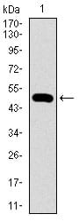
Figure 1: Western blot analysis using CTNNBL1 mAb against human CTNNBL1 (AA: 390-557) recombinant protein. (Expected MW is 45.8 kDa)
Western Blot
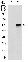
Figure 2: Western blot analysis using CTNNBL1 mAb against HEK293 (1) and CTNNBL1 (AA: 390-557)-hIgGFc transfected HEK293 (2) cell lysate.
Western Blot
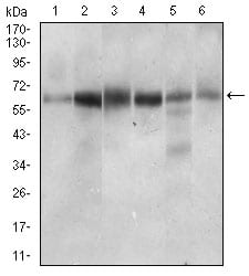
Figure 3: Western blot analysis using CTNNBL1 mouse mAb against Hela (1), Jurkat (2), HEK293 (3), A431 (4), HepG2 (5), RAJI (6) cell lysate.
Immunofluorescence analysis
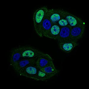
Figure 4: Immunofluorescence analysis of MCF-7 cells using CTNNBL1 mouse mAb (green). Blue: DRAQ5 fluorescent DNA dye. Secondary antibody from Fisher (Cat#: 35503)
Flow cytometric

Figure 5: Flow cytometric analysis of Hela cells using CTNNBL1 mouse mAb (green) and negative control (red).
Immunohistochemical analysis
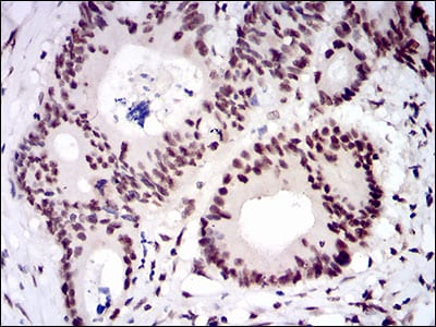
Figure 6: Immunohistochemical analysis of paraffin-embedded colon cancer tissues using CTNNBL1 mouse mAb with DAB staining.
Immunohistochemical analysis
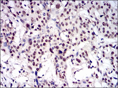
Figure 7: Immunohistochemical analysis of paraffin-embedded esophageal cancer tissues using CTNNBL1 mouse mAb with DAB staining.
Elisa
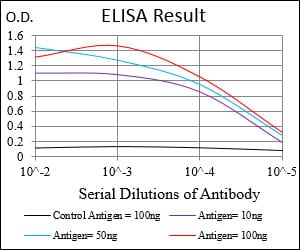
Black line: Control Antigen (100 ng); Purple line: Antigen(10ng); Blue line: Antigen (50 ng); Red line: Antigen (100 ng);
For Research Use Only. Not for use in diagnostic procedures.