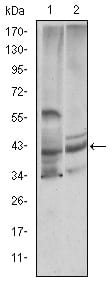
CST3 Primary Antibody
The cystatin superfamily encompasses proteins that contain multiple cystatin-like sequences. Some of the members are active cysteine protease inhibitors, while others have lost or perhaps never acquired this inhibitory activity. There are three inhibitory families in the superfamily, including the type 1 cystatins (stefins), type 2 cystatins and the kininogens. The type 2 cystatin proteins are a class of cysteine proteinase inhibitors found in a variety of human fluids and secretions, where they appear to provide protective functions. The cystatin locus on chromosome 20 contains the majority of the type 2 cystatin genes and pseudogenes. This gene is located in the cystatin locus and encodes the most abundant extracellular inhibitor of cysteine proteases, which is found in high concentrations in biological fluids and is expressed in virtually all organs of the body. A mutation in this gene has been associated with amyloid angiopathy. Expression of this protein in vascular wall smooth muscle cells is severely reduced in both atherosclerotic and aneurysmal aortic lesions, establishing its role in vascular disease. (provided by RefSeq)Tissue specificity: Expressed in submandibular and sublingual saliva but not in parotid saliva (at protein level). Expressed in various body fluids, such as the cerebrospinal fluid and plasma. Expressed in highest levels in the epididymis, vas deferens, brain, thymus, and ovary and the lowest in the submandibular gland.
2. Clin J Am Soc Nephrol. 2008 Nov;3(6):1610-4.