

CSNK2B Primary Antibody
Item Information
Catalog #
Size
Price
Description
This gene encodes the beta subunit of casein kinase II, a ubiquitous protein kinase which regulates metabolic pathways, signal transduction, transcription, translation, and replication. The enzyme is composed of three subunits, alpha, alpha prime and beta, which form a tetrameric holoenzyme. The alpha and alpha prime subunits are catalytic, while the beta subunit serves regulatory functions. The enzyme localizes to the endoplasmic reticulum and the Golgi apparatus. Two transcript variants encoding different isoforms have been found for this gene.
Product Overview
Entrez GenelD
1460
Aliases
G5A; CK2B; CK2N; CSK2B
Clone#
2F12F3
Host / Isotype
Mouse / IgG1
Species Reactivity
Human
Immunogen
Purified recombinant fragment of human CSNK2B (AA: FULL(1-215)) expressed in E. Coli.
Formulation
Purified antibody from tissue culture in PBS with 0.05% sodium azide
Storage
Store at 4°C short term. Aliquot and store at -20°C long term. Avoid freeze/thaw cycles.
Product Applications
WB (Western Blot)
1/500 - 1/2000
IHC_P(Immunohistochemistry)
1/200 - 1/1000
ICC (Immunocytochemistry)
1/200 - 1/1000
FCM (Flow Cytometry)
1/200 - 1/400
ELISA
1/10000
References
1. Nan Fang Yi Ke Da Xue Xue Bao. 2012 Oct;32(10):1491-4.
2. Am J Pathol. 2009 Jan;174(1):287-96.
2. Am J Pathol. 2009 Jan;174(1):287-96.
Product Image
Western Blot
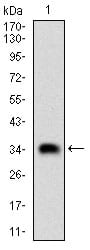
Figure 1: Western blot analysis using CSNK2B mAb against human CSNK2B (AA: FULL(1-215)) recombinant protein. (Expected MW is 35 kDa)
Western Blot
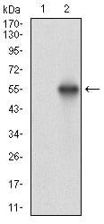
Figure 2: Western blot analysis using CSNK2B mAb against HEK293 (1) and CSNK2B (AA: FULL(1-215))-hIgGFc transfected HEK293 (2) cell lysate.
Western Blot
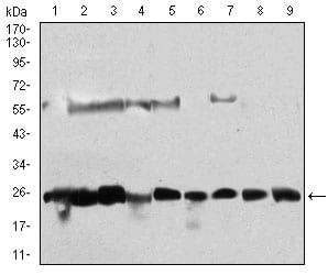
Figure 3: Western blot analysis using CSNK2B mouse mAb against Hela (1), Jurkat (2), K562 (3), HepG2 (4), C6 (5), SK-N-SH (6), NTERA-2 (7), MCF-7 (8), NIH/3T3 (9) cell lysate.
Immunofluorescence analysis
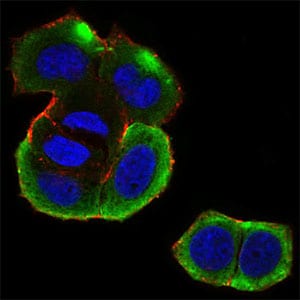
Figure 4: Immunofluorescence analysis of MCF-7 cells using CSNK2B mouse mAb (green). Blue: DRAQ5 fluorescent DNA dye. Red: Actin filaments have been labeled with Alexa Fluor-555 phalloidin. Secondary antibody from Fisher (Cat#: 35503)
Flow cytometric
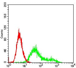
Figure 5: Flow cytometric analysis of Hela cells using CSNK2B mouse mAb (green) and negative control (red).
Immunohistochemical analysis

Figure 6: Immunohistochemical analysis of paraffin-embedded cervical cancer tissues using CSNK2B mouse mAb with DAB staining.
Immunohistochemical analysis
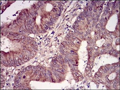
Figure 7: Immunohistochemical analysis of paraffin-embedded colon cancer tissues using CSNK2B mouse mAb with DAB staining.
Elisa
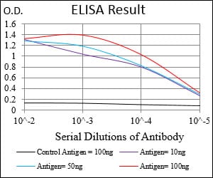
Black line: Control Antigen (100 ng); Purple line: Antigen(10ng); Blue line: Antigen (50 ng); Red line: Antigen (100 ng);
For Research Use Only. Not for use in diagnostic procedures.