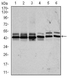
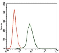
CSNK2A2 Primary Antibody

Item Information
Catalog #
Size
Price
Description

Casein kinase II (CK2) is a constitutively active, ubiquitously expressed serine/threonine protein kinase that is thought to have a regulatory function in cell proliferation, cell differentiation and apoptosis. CK2 functions as a tetrameric complex consisting of two regulatory beta-subunits and two catalytic units (alpha and alpha') in a homomeric or heteromeric conformation. Whilst the alpha- and alpha'-subunits are catalytically identical, proteins that regulate CK2, such as cdc2 and Hsp90, preferentially bind to the alpha and not the alpha'-subunit. CK2 can phosphorylate a number of key intracellular signaling proteins implicated in tumor suppression (p53 and PTEN) and tumorigenesis (myc, jun, NF-kappaB). CK2 is also thought to influence Wnt signaling via beta-catenin phosphorylation and the PI 3-K signaling pathway via the phosphorylation of Akt.

Product Overview
Entrez GenelD
1459
Aliases
CK2A2; CSNK2A1; FLJ43934
Clone#
1E1
Host / Isotype
Mouse / IgG1
Species Reactivity
Human, Mouse
Immunogen
Purified recombinant fragment of human CSNK2A2 (AA: 194-350) expressed in E. Coli.
Formulation
Ascitic fluid containing 0.03% sodium azide.
Storage
4°C; -20°C for long term storage
Product Applications
WB (Western Blot)
1/500 - 1/2000
FCM (Flow Cytometry)
1/200 - 1/400
ELISA
1/10000
References
1.Eur J Immunol. 2009 Jan;39(1):267-79. 2.Cell Mol Life Sci. 2009 Jan;66(2):339-49.
Product Image
Western Blot
Figure 1: Western blot analysis using CSNK2A2 mAb against human CSNK2A2 recombinant protein. (Expected MW is 44 kDa)
Western Blot
Figure 2: Western blot analysis using CSNK2A2 mouse mAb against HeLa (1), MCF-7 (2), HepG2 (3), Jurkat (4), NIH3T3 (5), and PC-12 (6) cell lysate.
Flow cytometric
Figure 3: Flow cytometric analysis of HepG2 cells using CSNK2A2 mouse mAb (green) and negative control (red).
Elisa
Black line: Control Antigen (100 ng); Purple line: Antigen(10ng); Blue line: Antigen (50 ng); Red line: Antigen (100 ng);
For Research Use Only. Not for use in diagnostic procedures.