

CSK Primary Antibody
Item Information
Catalog #
Size
Price
Description
Carboxy-terminal Src kinase (Csk) is a ubiquitously expressed nonreceptor tyrosine kinase that negatively regulates the Src family kinases (SFK) by phosphorylation of the SFK carboxy-terminal tyrosine. Phosphorylated carboxy-terminal tyrosine binds to the SH2 domain of SFK intramolecularly and leads to folding and inactivation of the SFK . This Csk-catalyzed SFK tyrosine phosphorylation is highly specific and exclusive. The SFK carboxy-terminal tyrosine is the only known physiological substrate of Csk .Tissue specificity: Expressed in lung and macrophages.
Product Overview
Entrez GenelD
1445
Aliases
MGC117393; CSK
Clone#
5F3
Host / Isotype
Mouse / IgG1
Species Reactivity
Human, Mouse, Monkey, Rat
Immunogen
Purified recombinant fragment of human CSK expressed in E. Coli.
Formulation
Ascitic fluid containing 0.03% sodium azide.
Storage
Store at 4°C short term. Aliquot and store at -20°C long term. Avoid freeze/thaw cycles.
Product Applications
WB (Western Blot)
1/500 - 1/2000
ICC (Immunocytochemistry)
1/200 - 1/1000
FCM (Flow Cytometry)
1/200 - 1/400
ELISA
1/10000
References
1. Nat Genet. 2009 Jun;41(6):677-87.
2. Leuk Res. 2009 Sep;33(9):e168-9.
3. J Hypertens. 2011 Jan;29(1):62-9.
2. Leuk Res. 2009 Sep;33(9):e168-9.
3. J Hypertens. 2011 Jan;29(1):62-9.
Product Image
Western Blot

Figure 1: Western blot analysis using CSK mouse mAb against NIH/3T3 (1)?Hela (2)?COS7 (3), Jurkat (4), Raw246.7 (5), A549 (6), HL-60 (7) and PC-12 (8) cell lysate.
Immunofluorescence analysis
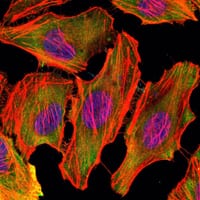
Figure 2: Immunofluorescence analysis of U251 cells using CSK mouse mAb (green). Blue: DRAQ5 fluorescent DNA dye. Red: Actin filaments have been labeled with Alexa Fluor-555 phalloidin.
Flow cytometric

Figure 3: Flow cytometric analysis of HL-60 cells using CSK mouse mAb (green) and negative control (purple).
For Research Use Only. Not for use in diagnostic procedures.