

CSF3 Primary Antibody
Item Information
Catalog #
Size
Price
Description
The protein encoded by this gene is a cytokine that controls the production, differentiation, and function of granulocytes. The active protein is found extracellularly. Alternatively spliced transcript variants have been described for this gene. [provided by RefSeq, May 2010]
Product Overview
Entrez GenelD
1440
Aliases
GCSF; CSF3OS; C17orf33
Clone#
8G5F7
Host / Isotype
Mouse / IgG1
Species Reactivity
Human
Immunogen
Purified recombinant fragment of human CSF3 (AA: 1-207) expressed in E. Coli.
Formulation
Purified antibody from tissue culture in PBS with 0.05% sodium azide
Storage
Store at 4°C short term. Aliquot and store at -20°C long term. Avoid freeze/thaw cycles.
Product Applications
WB (Western Blot)
1/500 - 1/2000
ICC (Immunocytochemistry)
1/200 - 1/1000
FCM (Flow Cytometry)
1/200 - 1/400
ELISA
1/10000
References
1.Biomarkers. 2012 Jun;17(4):319-24.
2.Eur J Immunol. 2010 Nov;40(11):3097-106.
2.Eur J Immunol. 2010 Nov;40(11):3097-106.
Product Image
Elisa
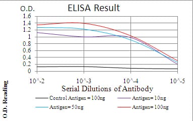
Figure 1: Black line: Control Antigen (100 ng); Purple line: Antigen(10ng); Blue line: Antigen (50 ng); Red line: Antigen (100 ng);
Western Blot
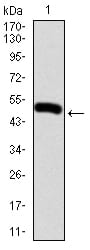
Figure 2:Western blot analysis using CSF3 mAb against human CSF3 (AA: 1-207) recombinant protein. (Expected MW is 48.2 kDa)
Western Blot
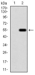
Figure 3:Western blot analysis using CSF3 mAb against HEK293 (1) and CSF3 (AA: 1-207)-hIgGFc transfected HEK293 (2) cell lysate.
Immunofluorescence analysis
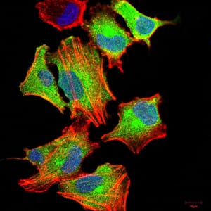
Figure 4:Immunofluorescence analysis of A549 cells using CSF3 mouse mAb (green). Blue: DRAQ5 fluorescent DNA dye. Red: Actin filaments have been labeled with Alexa Fluor- 555 phalloidin. Secondary antibody from Fisher (Cat#: 35503)
Immunofluorescence analysis

Figure 5:Immunofluorescence analysis of Hela cells using CSF3 mouse mAb (green). Blue: DRAQ5 fluorescent DNA dye. Red: Actin filaments have been labeled with Alexa Fluor- 555 phalloidin. Secondary antibody from Fisher (Cat#: 35503)
Flow cytometric
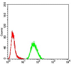
Figure 6:Flow cytometric analysis of A549 cells using CSF3 mouse mAb (green) and negative control (red).
For Research Use Only. Not for use in diagnostic procedures.