

CSF1R Primary Antibody
Item Information
Catalog #
Size
Price
Description
The protein encoded by this gene is the receptor for colony stimulating factor 1, a cytokine which controls the production, differentiation, and function of macrophages. This receptor mediates most if not all of the biological effects of this cytokine. Ligand binding activates the receptor kinase through a process of oligomerization and transphosphorylation. The encoded protein is a tyrosine kinase transmembrane receptor and member of the CSF1/PDGF receptor family of tyrosine-protein kinases. Mutations in this gene have been associated with a predisposition to myeloid malignancy. The first intron of this gene contains a transcriptionally inactive ribosomal protein L7 processed pseudogene oriented in the opposite direction.
Product Overview
Entrez GenelD
1436
Aliases
FMS; CSFR; FIM2; HDLS; C-FMS; CD115; CSF-1R; M-CSF-R
Clone#
4C9G2
Host / Isotype
Mouse / IgG1
Species Reactivity
Human
Immunogen
Purified recombinant fragment of human CSF1R (AA: 20-152) expressed in E. Coli.
Formulation
Purified antibody in PBS with 0.05% sodium azide
Storage
Store at 4°C short term. Aliquot and store at -20°C long term. Avoid freeze/thaw cycles.
Product Applications
WB (Western Blot)
1/500 - 1/2000
FCM (Flow Cytometry)
1/200 - 1/400
ELISA
1/10000
References
PLoS One. 2011;6(11):e27450.
J Biochem. 2012 Jan;151(1):47-55.
J Biochem. 2012 Jan;151(1):47-55.
Product Image
Elisa

Figure 1: Black line: Control Antigen (100 ng); Purple line: Antigen(10ng); Blue line: Antigen (50 ng); Red line: Antigen (100 ng);
Western Blot
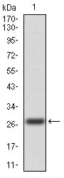
Figure 2:Western blot analysis using CSF1R mAb against human CSF1R (AA: 20-152) recombinant protein. (Expected MW is 27.2 kDa)
Western Blot

Figure 3:Western blot analysis using CSF1R mAb against HEK293 (1) and CSF1R (AA: 20-152)-hIgGFc transfected HEK293 (2) cell lysate.
Flow cytometric

Figure 4:Flow cytometric analysis of Hela cells using CSF1R mouse mAb (green) and negative control (red).
For Research Use Only. Not for use in diagnostic procedures.