
CRP Primary Antibody
Item Information
Catalog #
Size
Price
Description
The protein encoded by this gene belongs to the pentaxin family. It is involved in several host defense related functions based on its ability to recognize foreign pathogens and damaged cells of the host and to initiate their elimination by interacting with humoral and cellular effector systems in the blood. Consequently, the level of this protein in plasma increases greatly during acute phase response to tissue injury, infection, or other inflammatory stimuli.
Product Overview
Entrez GenelD
1401
Aliases
PTX1
Clone#
1G1
Host / Isotype
Mouse / IgG1
Species Reactivity
Human
Immunogen
Purified recombinant fragment of human CRP expressed in E. Coli.
Formulation
Ascitic fluid containing 0.03% sodium azide.
Storage
Store at 4°C short term. Aliquot and store at -20°C long term. Avoid freeze/thaw cycles.
Product Applications
WB (Western Blot)
1/500 - 1/2000
IHC_P(Immunohistochemistry)
1/200 - 1/1000
FCM (Flow Cytometry)
1/200 - 1/400
ELISA
1/10000
References
1. Clin Cardiol. 2010 Nov;33(11):708-14.
2. Mol Cell Biochem. 2011 Jan;347(1-2):183-9.
2. Mol Cell Biochem. 2011 Jan;347(1-2):183-9.
Product Image
Western Blot

Figure 1: Western blot analysis using CRP mAb against human CRP recombinant protein. ( AA: 1-224, Expected MW is 51 kDa)
Flow cytometric
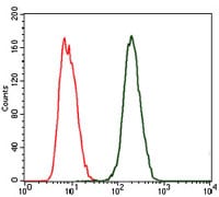
Figure 2: Flow cytometric analysis of MCF-7 cells using CRP mouse mAb (green) and negative control (red).
Immunohistochemical analysis

Figure 3: Immunohistochemical analysis of paraffin-embedded liver cancer tissues using CRP mouse mAb with DAB staining.
Elisa
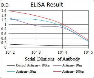
Black line: Control Antigen (100 ng); Purple line: Antigen(10ng); Blue line: Antigen (50 ng); Red line: Antigen (100 ng);
For Research Use Only. Not for use in diagnostic procedures.