

CRK Primary Antibody
Item Information
Catalog #
Size
Price
Description
This gene encodes a member of an adapter protein family that binds to several tyrosine-phosphorylated proteins. The product of this gene has several SH2 and SH3 domains (src-homology domains) and is involved in several signaling pathways, recruiting cytoplasmic proteins in the vicinity of tyrosine kinase through SH2-phosphotyrosine interaction. The N-terminal SH2 domain of this protein functions as a positive regulator of transformation whereas the C-terminal SH3 domain functions as a negative regulator of transformation. Two alternative transcripts encoding different isoforms with distinct biological activity have been described.
Product Overview
Entrez GenelD
1398
Aliases
CRKII
Clone#
3G11C1
Host / Isotype
Mouse / IgG2b
Species Reactivity
Human
Immunogen
Purified recombinant fragment of human CRK expressed in E. Coli.
Formulation
Ascitic fluid containing 0.03% sodium azide.
Storage
Store at 4°C short term. Aliquot and store at -20°C long term. Avoid freeze/thaw cycles.
Product Applications
WB (Western Blot)
1/500 - 1/2000
IHC_P(Immunohistochemistry)
1/200 - 1/1000
ICC (Immunocytochemistry)
1/200 - 1/1000
FCM (Flow Cytometry)
1/200 - 1/400
ELISA
1/10000
References
1. Seikagaku. 2009 May;81(5):361-76.
2. Mol Cancer Res. 2009 Sep;7(9):1582-92.
2. Mol Cancer Res. 2009 Sep;7(9):1582-92.
Product Image
Western Blot

Figure 1: Western blot analysis using CRK mAb against human CRK (AA: 1-204) recombinant protein. (Expected MW is 48.4 kDa)
Western Blot

Figure 2: Western blot analysis using CRK mAb against HEK293 (1) and CRK(AA: 1-204)-hIgGFc transfected HEK293 (2) cell lysate.
Immunohistochemical analysis

Figure 3: Immunohistochemical analysis of paraffin-embedded rectum cancer tissues using CRK mouse mAb with DAB staining.
Immunohistochemical analysis
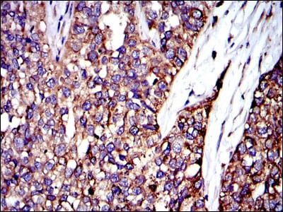
Figure 4: Immunohistochemical analysis of paraffin-embedded bladder cancer tissues using CRK mouse mAb with DAB staining.
Immunofluorescence analysis

Figure 5: Immunofluorescence analysis of 3T3-L1 cells using CRK mouse mAb (green). Blue: DRAQ5 fluorescent DNA dye. Red: Actin filaments have been labeled with Alexa Fluor-555 phalloidin.
Flow cytometric

Figure 6: Flow cytometric analysis of MCF-7 cells using CRK mouse mAb (blue) and negative control (red).
Elisa

Red: Control Antigen (100ng); Purple: Antigen (10ng); Green: Antigen (50ng); Blue: Antigen (100ng);
For Research Use Only. Not for use in diagnostic procedures.