

COX4I1 Primary Antibody
Item Information
Catalog #
Size
Price
Description
Cytochrome c oxidase (COX) functions as the terminal oxidase of the respiratory chain that uses cytochrome c as an electron donor to drive a proton gradient across the inner mitochondrial membrane. The mammalian COX apoenzyme is a heteromer consisting of three mitochondrial encoded catalytic subunits and several nuclear gene encoded structural subunits. COX contains two iron-coordination sites and two copper-coordination sites. Cytochrome c oxidase IV (COX4) is a nuclear-encoded subunit of COX that may play a role in regulating COX activity. COX4 is expressed ubiquitously in adult human tissue with the strongest levels of expression in the pancreas and moderate expression levels in heart, skeletal muscle and placenta.
Product Overview
Entrez GenelD
1327
Aliases
COX4; COXIV; COX4-1; MGC72016; COX4I1
Clone#
6B3
Host / Isotype
Mouse / IgG1
Species Reactivity
Human, Monkey, Rat, Mouse
Immunogen
Purified recombinant fragment of human COX4I1 expressed in E. Coli.
Formulation
Ascitic fluid containing 0.03% sodium azide.
Storage
Store at 4°C short term. Aliquot and store at -20°C long term. Avoid freeze/thaw cycles.
Product Applications
WB (Western Blot)
1/500 - 1/2000
ICC (Immunocytochemistry)
1/200 - 1/1000
FCM (Flow Cytometry)
1/200 - 1/400
ELISA
1/10000
References
1. Biochim Biophys Acta. 1992 Feb 26;1119(2):218-24.
2. Histochemistry. 1990;94(2):211-5.
3. FEBS Lett. 2000 Jun 30;476(1-2):22-6.
2. Histochemistry. 1990;94(2):211-5.
3. FEBS Lett. 2000 Jun 30;476(1-2):22-6.
Product Image
Western Blot
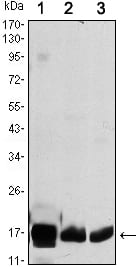
Figure 1: Western blot analysis using COX4I1 mouse mAb against HEK293 (1), A549 (2) and PC12 (3) cell lysate.
Immunofluorescence analysis

Figure 2: Immunofluorescence analysis of PANC-1 cells using COX4I1 mouse mAb (green). Blue: DRAQ5 fluorescent DNA dye. Red: Actin filaments have been labeled with Alexa Fluor-555 phalloidin.
Flow cytometric

Figure 3: Flow cytometric analysis of K562 cells using COX4I1 mouse mAb (blue) and negative control (red).
For Research Use Only. Not for use in diagnostic procedures.