

COL2A1 Primary Antibody
Item Information
Catalog #
Size
Price
Description
This gene encodes the alpha-1 chain of type II collagen, a fibrillar collagen found in cartilage and the vitreous humor of the eye. Mutations in this gene are associated with achondrogenesis, chondrodysplasia, early onset familial osteoarthritis, SED congenita, Langer-Saldino achondrogenesis, Kniest dysplasia, Stickler syndrome type I, and spondyloepimetaphyseal dysplasia Strudwick type. In addition, defects in processing chondrocalcin, a calcium binding protein that is the C-propeptide of this collagen molecule, are also associated with chondrodysplasia. There are two transcripts identified for this gene.
Product Overview
Entrez GenelD
1280
Aliases
AOM; ANFH; SEDC; STL1; COL11A3
Clone#
7G2C8
Host / Isotype
Mouse / Mouse IgG1
Species Reactivity
Human
Immunogen
Purified recombinant fragment of human COL2A1 (AA: 1222-1487) expressed in HEK293-6e cells supernatant.
Formulation
Purified antibody in PBS with 0.05% sodium azide
Storage
Store at 4°C short term. Aliquot and store at -20°C long term. Avoid freeze/thaw cycles.
Product Applications
WB (Western Blot)
1/500 - 1/2000
ICC (Immunocytochemistry)
1/50 - 1/200
FCM (Flow Cytometry)
1/200 - 1/400
ELISA
1/10000
References
1.J Clin Pediatr Dent. 2020 Sep 1;44(5):364-372.
2.Int J Biol Sci. 2020 Jan 16;16(5):859-868.
2.Int J Biol Sci. 2020 Jan 16;16(5):859-868.
Product Image
Elisa
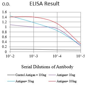
Figure 1:Black line: Control Antigen (100 ng);Purple line: Antigen (10ng); Blue line: Antigen (50 ng); Red line:Antigen (100 ng)
Western Blot
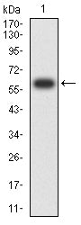
Figure 2:Western blot analysis using COL2A1 mAb against human COL2A1 (AA: 1222-1487) recombinant protein. (Expected MW is 60 kDa)
Western Blot

Figure 3:Western blot analysis using COL2A1 mouse mAb against Hela (1), MCF-7 (2),A549 (3),Jurkat (4), and K562 (5) cell lysate.
Immunohistochemical analysis

Figure 4:Immunofluorescence analysis of Hela cells using COL2A1 mouse mAb (green). Blue: DRAQ5 fluorescent DNA dye. Red: Actin filaments have been labeled with Alexa Fluor- 555 phalloidin. Secondary antibody from Fisher (Cat#: 35503)
Immunofluorescence analysis

Figure 5:Flow cytometric analysis of Jurkat cells using COL2A1 mouse mAb (green) and negative control (red).
For Research Use Only. Not for use in diagnostic procedures.