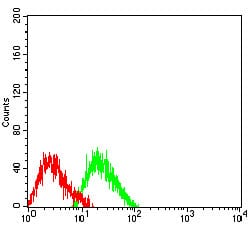
COL1A2 Primary Antibody

Item Information
Catalog #
Size
Price
Description

This gene encodes the pro-alpha2 chain of type I collagen whose triple helix comprises two alpha1 chains and one alpha2 chain. Type I is a fibril-forming collagen found in most connective tissues and is abundant in bone, cornea, dermis and tendon. Mutations in this gene are associated with osteogenesis imperfecta types I-IV, Ehlers-Danlos syndrome type VIIB, recessive Ehlers-Danlos syndrome Classical type, idiopathic osteoporosis, and atypical Marfan syndrome. Symptoms associated with mutations in this gene, however, tend to be less severe than mutations in the gene for the alpha1 chain of type I collagen (COL1A1) reflecting the different role of alpha2 chains in matrix integrity. Three transcripts, resulting from the use of alternate polyadenylation signals, have been identified for this gene.

Product Overview
Entrez GenelD
1278
Aliases
OI4; EDSCV; EDSARTH2
Clone#
4D1A7
Host / Isotype
Mouse / Mouse IgG1
Immunogen
Purified recombinant fragment of human COL1A2 (AA: 23-79) expressed in E. Coli.
Formulation
Purified antibody in PBS with 0.05% sodium azide
Storage
4?; -20? for long term storage
Product Applications
WB (Western Blot)
1/500 - 1/2000
ICC (Immunocytochemistry)
1/200 - 1/1000
FCM (Flow Cytometry)
1/200 - 1/400
ELISA
1/10000
References
1.BMC Med Genet. 2017 Dec 4;18(1):143. 2.Interact Cardiovasc Thorac Surg. 2017 Nov 1;25(5):813-817.
Product Image
Elisa
Figure 1:Black line: Control Antigen (100 ng);Purple line: Antigen (10ng); Blue line: Antigen (50 ng); Red line:Antigen (100 ng)
Western Blot
Figure 2:Western blot analysis using COL1A2 mAb against human COL1A2 (AA: 23-79) recombinant protein. (Expected MW is 31.4 kDa)
Western Blot
Figure 3:Western blot analysis using COL1A2 mAb against HEK293 (1) and COL1A2 (AA: 23-79)-hIgGFc transfected HEK293 (2) cell lysate.
Immunofluorescence analysis
Figure 4:Immunofluorescence analysis of Hela cells using COL1A2 mouse mAb (green). Blue: DRAQ5 fluorescent DNA dye. Red: Actin filaments have been labeled with Alexa Fluor- 555 phalloidin. Secondary antibody from Fisher (Cat#: 35503)
Flow cytometric
Figure 5:Flow cytometric analysis of SW480 cells using COL1A2 mouse mAb (green) and negative control (red).
For Research Use Only. Not for use in diagnostic procedures.