

CNN3 Primary Antibody
Item Information
Catalog #
Size
Price
Description
This gene encodes a protein with a markedly acidic C terminus; the basic N-terminus is highly homologous to the N-terminus of a related gene, CNN1. Members of the CNN gene family all contain similar tandemly repeated motifs. This encoded protein is associated with the cytoskeleton but is not involved in contraction.
Product Overview
Entrez GenelD
1266
Clone#
5E4E4
Host / Isotype
Mouse / Mouse IgG1
Immunogen
Purified recombinant fragment of human CNN3 (AA: 26-130) expressed in E. Coli.
Formulation
Purified antibody in PBS with 0.05% sodium azide
Storage
Store at 4°C short term. Aliquot and store at -20°C long term. Avoid freeze/thaw cycles.
Product Applications
WB (Western Blot)
1/500 - 1/2000
IHC_P(Immunohistochemistry)
1/200 - 1/1000
ICC (Immunocytochemistry)
1/500 - 1/2000
FCM (Flow Cytometry)
1/200 - 1/400
ELISA
1/10000
References
1.PLoS One. 2014 Jul 22;9(7):e103216. 2.Arch Dermatol Res. 2013 Sep;305(7):571-84.
Product Image
Elisa
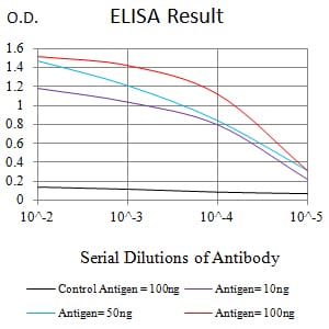
Figure 1:Black line: Control Antigen (100 ng);Purple line: Antigen (10ng); Blue line: Antigen (50 ng); Red line:Antigen (100 ng)
Western Blot

Figure 2:Western blot analysis using CNN3 mAb against human CNN3 (AA: 26-130) recombinant protein. (Expected MW is 37.6 kDa)
Western Blot

Figure 3:Western blot analysis using CNN3 mAb against HEK293 (1) and CNN3 (AA: 26-130)-hIgGFc transfected HEK293 (2) cell lysate.
Western Blot

Figure 4:Western blot analysis using CNN3 mouse mAb against Hela (1), U251 (2), and HEK293 (3) cell lysate.
Immunofluorescence analysis

Figure 5:Immunofluorescence analysis of Hela cells using CNN3 mouse mAb (green). Blue: DRAQ5 fluorescent DNA dye. Red: Actin filaments have been labeled with Alexa Fluor- 555 phalloidin. Secondary antibody from Fisher (Cat#: 35503)
Flow Cytometric

Figure 6:Flow cytometric analysis of Hela cells using CNN3 mouse mAb (green) and negative control (red).
Immunohistochemical Analysis

Figure 7:Immunohistochemical analysis of paraffin-embedded cervical cancer tissues using CNN3 mouse mAb with DAB staining.
Immunohistochemical Analysis

Figure 8:Immunohistochemical analysis of paraffin-embedded rectum cancer tissues using CNN3 mouse mAb with DAB staining.
For Research Use Only. Not for use in diagnostic procedures.