CLDN6 Primary Antibody

Item Information
Catalog #
Size
Price
Description

Tight junctions represent one mode of cell-to-cell adhesion in epithelial or endothelial cell sheets, forming continuous seals around cells and serving as a physical barrier to prevent solutes and water from passing freely through the paracellular space. These junctions are comprised of sets of continuous networking strands in the outwardly facing cytoplasmic leaflet, with complementary grooves in the inwardly facing extracytoplasmic leaflet. This gene encodes a component of tight junction strands, which is a member of the claudin family. The protein is an integral membrane protein and is one of the entry cofactors for hepatitis C virus. The gene methylation may be involved in esophageal tumorigenesis. This gene is adjacent to another family member CLDN9 on chromosome 16.[provided by RefSeq, Aug 2010]
Product Overview
Entrez GenelD
9074
Aliases
CLDN6
Clone#
8G5D2
Host / Isotype
Mouse / Mouse IgG1
Immunogen
Purified recombinant fragment of human CLDN6 (AA: extra(29-81) and extra(138-160)) expressed in E. Coli.
Formulation
Purified antibody in PBS with 0.05% sodium azide
Storage
4°C; -20°C for long term storage
Product Applications
WB (Western Blot)
1/500 - 1/2000
FCM (Flow Cytometry)
1/200-1/400
ELISA
1/10000
References
1,Oncol Rep. 2017 Aug;38(2):875-885. 2,Biotechnol J. 2018 Mar;13(3):e1700345.
Product Image
ELISA
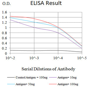
Figure 1: Black line: Control Antigen (100 ng);Purple line: Antigen (10ng); Blue line: Antigen (50 ng); Red line: Antigen (100 ng)
WESTERN BLOT
Figure 2: Western blot analysis using CLDN6 mAb against human CLDN6 (AA: extra(29-81) and extra(138-160)) recombinant protein. (Expected MW is 26 kDa)
WESTERN BLOT
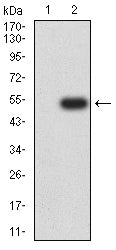
Figure 3: Western blot analysis using CLDN6 mAb against HEK293-6e (1) and CLDN6 (AA: extra(29-81) and extra(138-160))-hIgGFc transfected HEK293-6e (2) cell lysate.
FLOW CYTOMETRY
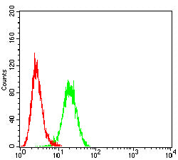
Figure 4: Flow cytometric analysis of Hepg2 cells using CLDN6 mouse mAb (green) and negative control (red).
For Research Use Only. Not for use in diagnostic procedures.