

CK5 Primary Antibody
Item Information
Catalog #
Size
Price
Description
The protein encoded by this gene is a member of the keratin gene family. The type II cytokeratins consist of basic or neutral proteins which are arranged in pairs of heterotypic keratin chains coexpressed during differentiation of simple and stratified epithelial tissues. This type II cytokeratin is specifically expressed in the basal layer of the epidermis with family member KRT14. Mutations in these genes have been associated with a complex of diseases termed epidermolysis bullosa simplex. The type II cytokeratins are clustered in a region of chromosome 12q12-q13.
Product Overview
Entrez GenelD
3852
Aliases
KRT5; K5; DDD; EBS2; KRT5A
Clone#
10C11E6
Host / Isotype
Mouse / IgG1
Species Reactivity
Human
Immunogen
Purified recombinant fragment of human CK5 (AA: 316-590) expressed in E. Coli.
Formulation
Purified antibody in PBS with 0.05% sodium azide
Storage
Store at 4°C short term. Aliquot and store at -20°C long term. Avoid freeze/thaw cycles.
Product Applications
WB (Western Blot)
1/500 - 1/2000
IHC_P(Immunohistochemistry)
1/200 - 1/1000
ELISA
1/10000
References
1. Mol Biol Cell. 2011 Nov;22(21):4068-78.
2. Am J Surg Pathol. 2009 Nov;33(11):1615-23.
2. Am J Surg Pathol. 2009 Nov;33(11):1615-23.
Product Image
Western Blot

Figure 1: Western blot analysis using CK5 mAb against human CK5 recombinant protein. (Expected MW is 47.8 kDa)
Western Blot

Figure 2: Western blot analysis using CK5 mouse mAb against A431 cell lysate.
Immunohistochemical analysis

Figure 3: Immunohistochemical analysis of paraffin-embedded bladder cancer tissues using CK5 mouse mAb with DAB staining.
Immunohistochemical analysis
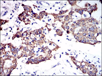
Figure 4: Immunohistochemical analysis of paraffin-embedded esophagus cancer tissues using CK5 mouse mAb with DAB staining.
Elisa

Black line: Control Antigen (100 ng); Purple line: Antigen(10ng); Blue line: Antigen (50 ng); Red line: Antigen (100 ng);
For Research Use Only. Not for use in diagnostic procedures.