

CHRNE Primary Antibody
Item Information
Catalog #
Size
Price
Description
Acetylcholine receptors at mature mammalian neuromuscular junctions are pentameric protein complexes composed of four subunits in the ratio of two alpha subunits to one beta, one epsilon, and one delta subunit. The acetylcholine receptor changes subunit composition shortly after birth when the epsilon subunit replaces the gamma subunit seen in embryonic receptors. Mutations in the epsilon subunit are associated with congenital myasthenic syndrome.
Product Overview
Entrez GenelD
1145
Aliases
ACHRE; CMS1D; CMS1E; CMS2A; CMS4A; CMS4B; CMS4C; FCCMS; SCCMS
Clone#
5F11G8
Host / Isotype
Mouse / IgG1
Species Reactivity
Human, Rat
Immunogen
Purified recombinant fragment of human CHRNE (AA: extra 21-239) expressed in E. Coli.
Formulation
Purified antibody in PBS with 0.05% sodium azide
Storage
Store at 4°C short term. Aliquot and store at -20°C long term. Avoid freeze/thaw cycles.
Product Applications
WB (Western Blot)
1/500 - 1/2000
FCM (Flow Cytometry)
1/200 - 1/400
ELISA
1/10000
References
1.J Neuroophthalmol. 2011 Mar;31(1):42-7.2.Neurology. 2008 Dec 9;71(24):1967-72.
Product Image
Elisa
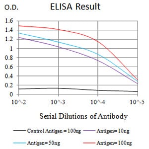
Figure 1: Black line: Control Antigen (100 ng);Purple line: Antigen (10ng); Blue line: Antigen (50 ng); Red line:Antigen (100 ng)
Western Blot
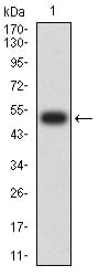
Figure 2:Western blot analysis using CHRNE mAb against human CHRNE (AA: extra 21-239) recombinant protein. (Expected MW is 50.1 kDa)
Western Blot
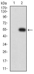
Figure 3:Western blot analysis using CHRNE mAb against HEK293 (1) and CHRNE (AA: extra 21-239)-hIgGFc transfected HEK293 (2) cell lysate.
Western Blot
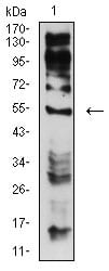
Figure 4:Western blot analysis using CHRNE mouse mAb against C6 (1) cell lysate.
Flow cytometric
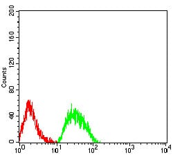
Figure 5:Flow cytometric analysis of SK-N-SH cells using CHRNE mouse mAb (green) and negative control (red).
For Research Use Only. Not for use in diagnostic procedures.