CHRNB3 Primary Antibody

Item Information
Catalog #
Size
Price
Description

The nicotinic acetylcholine receptors (nAChRs) are members of a superfamily of ligand-gated ion channels that mediate fast signal transmission at synapses. The nAChRs are (hetero)pentamers composed of homologous subunits. The subunits that make up the muscle and neuronal forms of nAChRs are encoded by separate genes and have different primary structure. There are several subtypes of neuronal nAChRs that vary based on which homologous subunits are arranged around the central channel. They are classified as alpha-subunits if, like muscle alpha-1 (MIM 100690), they have a pair of adjacent cysteines as part of the presumed acetylcholine binding site. Subunits lacking these cysteine residues are classified as beta-subunits (Groot Kormelink and Luyten, 1997 [PubMed 9009220]). Elliott et al. (1996) [PubMed 8906617] stated that the proposed structure for each subunit is a conserved N-terminal extracellular domain followed by 3 conserved transmembrane domains, a variable cytoplasmic loop, a fourth conserved transmembrane domain, and a short C-terminal extracellular region.
Product Overview
Entrez GenelD
1142
Clone#
7C9F12
Host / Isotype
Mouse / IgG1
Species Reactivity
Human
Immunogen
Purified recombinant fragment of human CHRNB3 (AA: extra 25-232) expressed in E. Coli.
Formulation
Purified antibody in PBS with 0.05% sodium azide
Storage
4°C; -20°C for long term storage
Product Applications
WB (Western Blot)
1/500 - 1/2000
FCM (Flow Cytometry)
1/200 - 1/400
ELISA
1/10000
References
1.Neurobiol Aging. 2014 Sep;35(9):2179.e1-6. 2.Mol Psychiatry. 2013 Nov;18(11):1149-51.
Product Image
Elisa
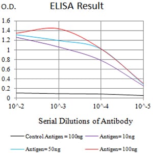
Figure 1: Black line: Control Antigen (100 ng);Purple line: Antigen (10ng); Blue line: Antigen (50 ng); Red line:Antigen (100 ng)
Western Blot
Figure 2:Western blot analysis using CHRNB3 mAb against human CHRNB3 (AA: extra 25-232) recombinant protein. (Expected MW is 50.2 kDa)
Western Blot
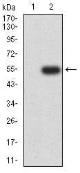
Figure 3:Western blot analysis using CHRNB3 mAb against HEK293 (1) and CHRNB3 (AA: extra 25-232)-hIgGFc transfected HEK293 (2) cell lysate.
Flow cytometric
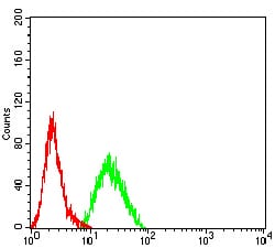
Figure 4:Flow cytometric analysis of SK-N-SH cells using CHRNB3 mouse mAb (green) and negative control (red).
For Research Use Only. Not for use in diagnostic procedures.