

CHRNA6 Primary Antibody
Item Information
Catalog #
Size
Price
Description
This gene encodes an alpha subunit of neuronal nicotinic acetylcholine receptors. These receptors consist of five subunits and function as ion channels involved in neurotransmission. The encoded protein is a subunit of neuronal nicotinic acetylcholine receptors that mediate dopaminergic neurotransmission and are activated by acetylcholine and exogenous nicotine. Alternatively spliced transcript variants have been observed for this gene. Single nucleotide polymorphisms in this gene have been associated with both nicotine and alcohol dependence.
Product Overview
Entrez GenelD
8973
Aliases
CHNRA6
Clone#
5B6G8
Host / Isotype
Mouse / IgG1
Species Reactivity
Human
Immunogen
Purified recombinant fragment of human CHRNA6 (AA: 26-239) expressed in E. Coli.
Formulation
Purified antibody in PBS with 0.05% sodium azide
Storage
Store at 4°C short term. Aliquot and store at -20°C long term. Avoid freeze/thaw cycles.
Product Applications
WB (Western Blot)
1/500 - 1/2000
IHC_P(Immunohistochemistry)
1/200 - 1/1000
ICC (Immunocytochemistry)
1/100 - 1/500
FCM (Flow Cytometry)
1/200 - 1/400
ELISA
1/10000
References
1.Mol Brain. 2014 May 2;7:35.
2.Mol Psychiatry. 2010 Jan;15(1):6-8.
2.Mol Psychiatry. 2010 Jan;15(1):6-8.
Product Image
Elisa
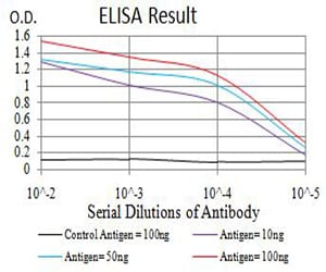
Figure 1: Black line: Control Antigen (100 ng);Purple line: Antigen (10ng); Blue line: Antigen (50 ng); Red line:Antigen (100 ng)
Western Blot
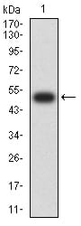
Figure 2:Western blot analysis using CHRNA6 mAb against human CHRNA6 (AA: 26-239) recombinant protein. (Expected MW is 51.2 kDa)
Western Blot
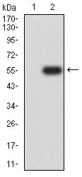
Figure 3:Western blot analysis using CHRNA6 mAb against HEK293 (1) and CHRNA6 (AA: 26-239)-hIgGFc transfected HEK293 (2) cell lysate.
Immunofluorescence analysis
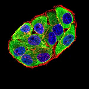
Figure 4:Immunofluorescence analysis of Hela cells using CHRNA6 mouse mAb (green). Blue: DRAQ5 fluorescent DNA dye. Red: Actin filaments have been labeled with Alexa Fluor- 555 phalloidin. Secondary antibody from Fisher (Cat#: 35503)
Flow cytometric
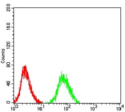
Figure 5:Flow cytometric analysis of SH-SY5Y cells using CHRNA6 mouse mAb (green) and negative control (red).
Flow cytometric
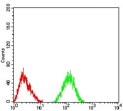
Figure 6:Flow cytometric analysis of SK-N-SH cells using CHRNA6 mouse mAb (green) and negative control (red).
Immunohistochemical analysis
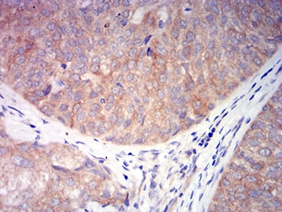
Figure 7:Immunohistochemical analysis of paraffin-embedded bladder cancer tissues using CHRNA6 mouse mAb with DAB staining.
For Research Use Only. Not for use in diagnostic procedures.