

CHRNA3 Primary Antibody
Item Information
Catalog #
Size
Price
Description
This locus encodes a member of the nicotinic acetylcholine receptor family of proteins. Members of this family of proteins form pentameric complexes comprised of both alpha and beta subunits. This locus encodes an alpha-type subunit, as it contains characteristic adjacent cysteine residues. The encoded protein is a ligand-gated ion channel that likely plays a role in neurotransmission. Polymorphisms in this gene have been associated with an increased risk of smoking initiation and an increased susceptibility to lung cancer. Alternatively spliced transcript variants have been described.
Product Overview
Entrez GenelD
1136
Aliases
LNCR2; PAOD2; NACHRA3
Clone#
6D3A10
Host / Isotype
Mouse / IgG1
Species Reactivity
Human
Immunogen
Purified recombinant fragment of human CHRNA3 (AA: 32-240) expressed in E. Coli.
Formulation
Purified antibody in PBS with 0.05% sodium azide
Storage
Store at 4°C short term. Aliquot and store at -20°C long term. Avoid freeze/thaw cycles.
Product Applications
WB (Western Blot)
1/500 - 1/2000
ICC (Immunocytochemistry)
1/200 - 1/1000
FCM (Flow Cytometry)
1/200 - 1/400
ELISA
1/10000
References
1.Tumour Biol. 2015 Jul;36(7):4987-92.
2.Int J Mol Sci. 2014 Mar 28;15(4):5446-57.
2.Int J Mol Sci. 2014 Mar 28;15(4):5446-57.
Product Image
Elisa
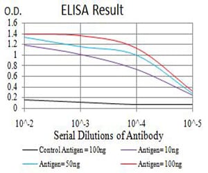
Figure 1: Black line: Control Antigen (100 ng);Purple line: Antigen (10ng); Blue line: Antigen (50 ng); Red line:Antigen (100 ng)
Western Blot

Figure 2:Western blot analysis using CHRNA3 mAb against human CHRNA3 (AA: 32-240) recombinant protein. (Expected MW is 50.6 kDa)
Western Blot

Figure 3:Western blot analysis using CHRNA3 mAb against HEK293 (1) and CHRNA3 (AA: 32-240)-hIgGFc transfected HEK293 (2) cell lysate.
Immunofluorescence analysis

Figure 4:Immunofluorescence analysis of Hela cells using CHRNA3 mouse mAb (green). Blue: DRAQ5 fluorescent DNA dye. Red: Actin filaments have been labeled with Alexa Fluor- 555 phalloidin. Secondary antibody from Fisher (Cat#: 35503)
Immunofluorescence analysis

Figure 5:Immunofluorescence analysis of SMMC-7721 cells using CHRNA3 mouse mAb (green). Blue: DRAQ5 fluorescent DNA dye. Red: Actin filaments have been labeled with Alexa Fluor- 555 phalloidin. Secondary antibody from Fisher (Cat#: 35503)
Flow cytometric

Figure 6:Flow cytometric analysis of SH-SY5Y cells using CHRNA3 mouse mAb (green) and negative control (red).
Flow cytometric

Figure 7:Flow cytometric analysis of SK-N-SH cells using CHRNA3 mouse mAb (green) and negative control (red).
For Research Use Only. Not for use in diagnostic procedures.