

CHD3 Primary Antibody
Item Information
Catalog #
Size
Price
Description
This gene encodes a member of the CHD family of proteins which are characterized by the presence of chromo (chromatin organization modifier) domains and SNF2-related helicase/ATPase domains. This protein is one of the components of a histone deacetylase complex referred to as the Mi-2/NuRD complex which participates in the remodeling of chromatin by deacetylating histones. Chromatin remodeling is essential for many processes including transcription. Autoantibodies against this protein are found in a subset of patients with dermatomyositis. Three alternatively spliced transcripts encoding different isoforms have been described.
Product Overview
Entrez GenelD
1107
Aliases
ZFH; Mi-2a; Mi2-ALPHA; CHD3
Clone#
2G4
Host / Isotype
Mouse / IgG1
Species Reactivity
Human, Mouse
Immunogen
Purified recombinant fragment of human CHD3 expressed in E. Coli.
Formulation
Purified antibody in PBS with 0.05% sodium azide.
Storage
Store at 4°C short term. Aliquot and store at -20°C long term. Avoid freeze/thaw cycles.
Product Applications
WB (Western Blot)
1/500 - 1/2000
IHC_P(Immunohistochemistry)
1/200 - 1/1000
ICC (Immunocytochemistry)
1/200 - 1/1000
FCM (Flow Cytometry)
1/200 - 1/400
ELISA
1/10000
References
1. Virus Res. 2003 Dec;98(1):83-91.
2. Mol Cell. 2004 Sep 24;15(6):853-65.
3. J Biol Chem. 2008 Dec 12;283(50):34976-82.
2. Mol Cell. 2004 Sep 24;15(6):853-65.
3. J Biol Chem. 2008 Dec 12;283(50):34976-82.
Product Image
Western Blot

Figure 1: Western blot analysis using CHD3 mouse mAb against Hela (1), K562 (2), Jurkat (3), NTERA-2 (4), HEK293 (5), Raji (6) cell lysate and mouse brain (7) tissue lysate.
Immunohistochemical analysis

Figure 2: Immunohistochemical analysis of paraffin-embedded colon cancer tissues using CHD3 mouse mAb with DAB staining.
Immunofluorescence analysis

Figure 3: Immunofluorescence analysis of Hela cells using CHD3 mouse mAb (green). Blue: DRAQ5 fluorescent DNA dye.
Flow cytometric
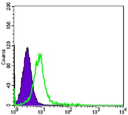
Figure 4: Flow cytometric analysis of K562 cells using CHD3 mouse mAb (green) and negative control (purple).
For Research Use Only. Not for use in diagnostic procedures.