

CFHR5 Primary Antibody
Item Information
Catalog #
Size
Price
Description
This gene is a member of a small complement factor H (CFH) gene cluster on chromosome 1. Each member of this gene family contains multiple short consensus repeats (SCRs) typical of regulators of complement activation. The protein encoded by this gene has nine SCRs with the first two repeats having heparin binding properties, a region within repeats 5-7 having heparin binding and C reactive protein binding properties, and the C-terminal repeats being similar to a complement component 3 b (C3b) binding domain. This protein co-localizes with C3, binds C3b in a dose-dependent manner, and is recruited to tissues damaged by C-reactive protein. Allelic variations in this gene have been associated, but not causally linked, with two different forms of kidney disease: membranoproliferative glomerulonephritis type II (MPGNII) and hemolytic uraemic syndrome (HUS).
Product Overview
Entrez GenelD
81494
Aliases
FHR5; CFHL5; FHR-5; CFHR5D
Clone#
3E1E10
Host / Isotype
Mouse / IgG1
Species Reactivity
Human
Immunogen
Purified recombinant fragment of human CFHR5 (AA: 344-569) expressed in E. Coli.
Formulation
Purified antibody in PBS with 0.05% sodium azide
Storage
Store at 4°C short term. Aliquot and store at -20°C long term. Avoid freeze/thaw cycles.
Product Applications
WB (Western Blot)
1/500 - 1/2000
IHC_P(Immunohistochemistry)
1/200 - 1/1000
ICC (Immunocytochemistry)
1/200 - 1/1000
FCM (Flow Cytometry)
1/200 - 1/400
ELISA
1/10000
References
1.Kidney Int. 2014 Apr;85(4):933-7.
2.Dis Model Mech. 2011 Nov;4(6):721-6.
2.Dis Model Mech. 2011 Nov;4(6):721-6.
Product Image
Elisa
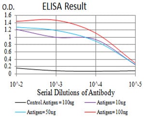
Figure 1: Black line: Control Antigen (100 ng);Purple line: Antigen (10ng); Blue line: Antigen (50 ng); Red line:Antigen (100 ng)
Western Blot
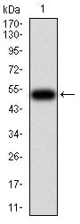
Figure 2:Western blot analysis using CFHR5 mAb against human CFHR5 (AA: 344-569) recombinant protein. (Expected MW is 51.8 kDa)
Western Blot
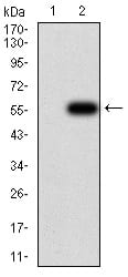
Figure 3:Western blot analysis using CFHR5 mAb against HEK293 (1) and CFHR5 (AA: 344-569)-hIgGFc transfected HEK293 (2) cell lysate.
Immunofluorescence analysis
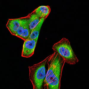
Figure 4:Immunofluorescence analysis of Hela cells using CFHR5 mouse mAb (green). Blue: DRAQ5 fluorescent DNA dye. Red: Actin filaments have been labeled with Alexa Fluor- 555 phalloidin. Secondary antibody from Fisher (Cat#: 35503)
Flow cytometric
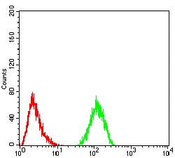
Figure 5:Flow cytometric analysis of Hela cells using CFHR5 mouse mAb (green) and negative control (red).
Immunohistochemical analysis
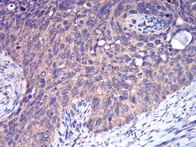
Figure 6:Immunohistochemical analysis of paraffin-embedded cervical cancer tissues using CFHR5 mouse mAb with DAB staining.
Immunohistochemical analysis
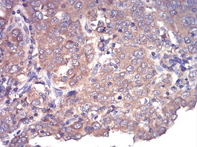
Figure 7:Immunohistochemical analysis of paraffin-embedded endometrial cancer tissues using CFHR5 mouse mAb with DAB staining.
For Research Use Only. Not for use in diagnostic procedures.