

CEBPA Primary Antibody
Item Information
Catalog #
Size
Price
Description
The protein encoded by this intronless gene is a bZIP transcription factor which can bind as a homodimer to certain promoters and enhancers. It can also form heterodimers with the related proteins CEBP-beta and CEBP-gamma. The encoded protein has been shown to bind to the promoter and modulate the expression of the gene encoding leptin, a protein that plays an important role in body weight homeostasis. Also, the encoded protein can interact with CDK2 and CDK4, thereby inhibiting these kinases and causing growth arrest in cultured cells.
Product Overview
Entrez GenelD
1050
Aliases
CEBP; C/EBP-alpha
Clone#
5B7
Host / Isotype
Mouse / IgG1
Species Reactivity
Human
Immunogen
Synthesized peptide of human CEBPA (AA: C-RKSRDKAKRNVETKV).
Formulation
Ascitic fluid containing 0.03% sodium azide.
Storage
Store at 4°C short term. Aliquot and store at -20°C long term. Avoid freeze/thaw cycles.
Product Applications
WB (Western Blot)
1/500 - 1/2000
IHC_P(Immunohistochemistry)
1/200 - 1/1000
ICC (Immunocytochemistry)
1/200 - 1/1000
FCM (Flow Cytometry)
1/200 - 1/400
ELISA
1/10000
References
Br J Cancer. 2010 Jul 13;103(2):275-84.
Cell Res. 2010 Apr;20(4):470-9.
Cell Res. 2010 Apr;20(4):470-9.
Product Image
Western Blot

Figure 1: Western blot analysis using CEBPA mouse mAb against Jurkat (1), k562 (2), and HepG2 (3) cell lysate.
Immunohistochemical analysis

Figure 2: Immunohistochemical analysis of paraffin-embedded rectum tissues using CEBPA mouse mAb with DAB staining.
Immunofluorescence analysis

Figure 3: Immunofluorescence analysis of HeLa cells. Blue: DRAQ5 fluorescent DNA dye. Red: Actin filaments have been labeled with Alexa Fluor-555 phalloidin.
Immunofluorescence analysis
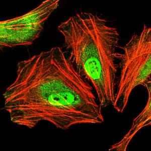
Figure 4: Immunofluorescence analysis of HeLa cells using CEBPA mouse mAb (green). Blue: DRAQ5 fluorescent DNA dye. Red: Actin filaments have been labeled with Alexa Fluor-555 phalloidin.
Flow cytometric

Figure 5: Flow cytometric analysis of MCF-7 cells using CEBPA mouse mAb (green) and negative control (red).
Elisa

Black line: Control Antigen (100 ng); Purple line: Antigen(10ng); Blue line: Antigen (50 ng); Red line: Antigen (100 ng);
For Research Use Only. Not for use in diagnostic procedures.