

CDX2 Primary Antibody
Item Information
Catalog #
Size
Price
Description
This gene is a member of the caudal-related homeobox transcription factor gene family. The encoded protein is a major regulator of intestine-specific genes involved in cell growth an differentiation. This protein also plays a role in early embryonic development of the intestinal tract. Aberrant expression of this gene is associated with intestinal inflammation and tumorigenesis. [provided by RefSeq, Jan 2012]
Product Overview
Entrez GenelD
1045
Aliases
CDX3; CDX-3; CDX2/AS
Clone#
5B4C1
Host / Isotype
Mouse / Mouse IgG1
Immunogen
Purified recombinant fragment of human CDX2 (AA: 1-180) expressed in E. Coli.
Formulation
Purified antibody in PBS with 0.05% sodium azide
Storage
Store at 4°C short term. Aliquot and store at -20°C long term. Avoid freeze/thaw cycles.
Product Applications
WB (Western Blot)
1/500 - 1/2000
IHC_P(Immunohistochemistry)
1/200-1/1000
FCM (Flow Cytometry)
1/200-1/400
ELISA
1/10000
References
1,Virchows Arch. 2020 Jul;477(1):21-31. 2,Am J Surg Pathol. 2019 Nov;43(11):1473-1482.
Product Image
ELISA
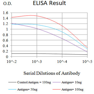
Figure 1: Black line: Control Antigen (100 ng);Purple line: Antigen (10ng); Blue line: Antigen (50 ng); Red line: Antigen (100 ng)
WESTERN BLOT
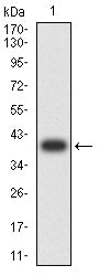
Figure 2: Western blot analysis using CDX2 mAb against human CDX2 (AA: 1-180) recombinant protein. (Expected MW is 40 kDa)
WESTERN BLOT
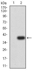
Figure 3: Western blot analysis using CDX2 mAb against HEK293-6e (1) and CDX2 (AA: 1-180)-hIgGFc transfected HEK293-6e (2) cell lysate.
FLOW CYTOMETRY
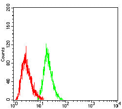
Figure 4: Flow cytometric analysis of THP-1 cells using CDX2 mouse mAb (green) and negative control (red).
FLOW CYTOMETRY
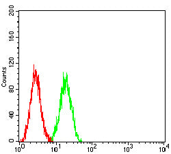
Figure 5: Flow cytometric analysis of SK-OV-3 cells using CDX2 mouse mAb (green) and negative control (red).
FLOW CYTOMETRY

Figure 6: Flow cytometric analysis of Jurkat cells using CDX2 mouse mAb (green) and negative control (red).
IMMUNOHISTOCHEMISTRY
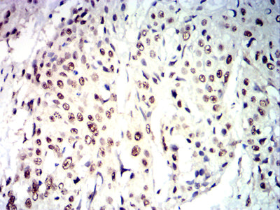
Figure 7: Immunohistochemical analysis of paraffin-embedded esophageal cancer tissues using CDX2 mouse mAb with DAB staining.
IMMUNOHISTOCHEMISTRY
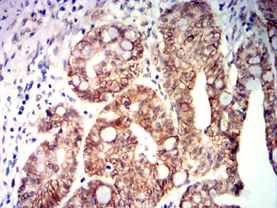
Figure 8: Immunohistochemical analysis of paraffin-embedded rectal cancer tissues using CDX2 mouse mAb with DAB staining.
For Research Use Only. Not for use in diagnostic procedures.