
CDK2 Primary Antibody
Item Information
Catalog #
Size
Price
Description
The protein encoded by this gene is a member of the Ser/Thr protein kinase family. This protein kinase is highly similar to the gene products of S. cerevisiae cdc28, and S. pombe cdc2. It is a catalytic subunit of the cyclin-dependent protein kinase complex, whose activity is restricted to the G1-S phase, and essential for cell cycle G1/S phase transition. This protein associates with and regulated by the regulatory subunits of the complex including cyclin A or E, CDK inhibitor p21Cip1 (CDKN1A) and p27Kip1 (CDKN1B). Its activity is also regulated by its protein phosphorylation. Two alternatively spliced variants and multiple transcription initiation sites of this gene have been reported.
Product Overview
Entrez GenelD
1017
Aliases
p33
Clone#
1A6
Host / Isotype
Mouse / IgG1
Species Reactivity
Human, Mouse
Immunogen
Purified recombinant fragment of human CDK2 expressed in E. Coli.
Formulation
Purified antibody in PBS with 0.05% sodium azide
Storage
Store at 4°C short term. Aliquot and store at -20°C long term. Avoid freeze/thaw cycles.
Product Applications
WB (Western Blot)
1/500 - 1/2000
IHC_P(Immunohistochemistry)
1/200 - 1/1000
ICC (Immunocytochemistry)
1/200 - 1/1000
FCM (Flow Cytometry)
1/200 - 1/400
ELISA
1/10000
References
Eur Arch Otorhinolaryngol. 2009 Oct;266(10):1501-7.
Cell Cycle. 2009 Jun 15;8(12):1952-63.
Cell Cycle. 2009 Jun 15;8(12):1952-63.
Product Image
Western Blot

Figure 1: Western blot analysis using CDK2 mAb against human CDK2 (AA: 197-295) recombinant protein. (Expected MW is 36.8 kDa)
Western Blot
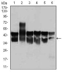
Figure 2: Western blot analysis using CDK2 mouse mAb against Jurkat (1), HL-60 (2), K562(3), A431(4), HeLa(5), and NIH3T3 (6) cell lysate.
Immunohistochemical analysis

Figure 3: Immunohistochemical analysis of paraffin-embedded cervical cancer tissues using CDK2 mouse mAb with DAB staining.
Immunohistochemical analysis
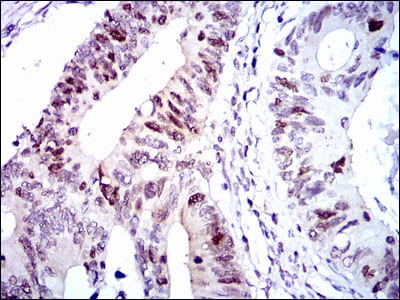
Figure 4: Immunohistochemical analysis of paraffin-embedded colon cancer tissues using CDK2 mouse mAb with DAB staining.
Immunofluorescence analysis

Figure 5: Immunofluorescence analysis of HeLa cells using CDK2 mouse mAb (green). Red: Actin filaments have been labeled with Alexa Fluor-555 phalloidin.
Flow cytometric

Figure 6: Flow cytometric analysis of Jurkat cells using CDK2 mouse mAb (green) and negative control (red).
Elisa

Black line: Control Antigen (100 ng); Purple line: Antigen(10ng); Blue line: Antigen (50 ng); Red line: Antigen (100 ng);
For Research Use Only. Not for use in diagnostic procedures.