

CDH17 Primary Antibody
Item Information
Catalog #
Size
Price
Description
This gene is a member of the cadherin superfamily, genes encoding calcium-dependent, membrane-associated glycoproteins. The encoded protein is cadherin-like, consisting of an extracellular region, containing 7 cadherin domains, and a transmembrane region but lacking the conserved cytoplasmic domain. The protein is a component of the gastrointestinal tract and pancreatic ducts, acting as an intestinal proton-dependent peptide transporter in the first step in oral absorption of many medically important peptide-based drugs. The protein may also play a role in the morphological organization of liver and intestine. Alternative splicing results in multiple transcript variants.
Product Overview
Entrez GenelD
1015
Aliases
HPT1; CDH16; HPT-1
Clone#
7D10E1
Host / Isotype
Mouse / IgG1
Species Reactivity
Human
Immunogen
Purified recombinant fragment of human CDH17 (AA: extra(600-707)) expressed in E. Coli.
Formulation
Purified antibody from tissue culture in PBS with 0.05% sodium azide
Storage
Store at 4°C short term. Aliquot and store at -20°C long term. Avoid freeze/thaw cycles.
Product Applications
WB (Western Blot)
1/500 - 1/2000
IHC_P(Immunohistochemistry)
1/200 - 1/1000
ELISA
1/10000
References
1. Cancer Biol Ther. 2013 Mar;14(3):262-70.
2. Mod Pathol. 2008 Nov;21(11):1379-86.
2. Mod Pathol. 2008 Nov;21(11):1379-86.
Product Image
Western Blot
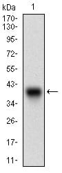
Figure 1: Western blot analysis using CDH17 mAb against human CDH17 (AA: extra(600-707)) recombinant protein. (Expected MW is 37.9 kDa)
Western Blot
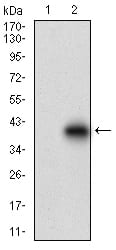
Figure 2: Western blot analysis using CDH17 mAb against HEK293 (1) and CDH17 (AA: extra(600-707))-hIgGFc transfected HEK293 (2) cell lysate.
Immunohistochemical analysis
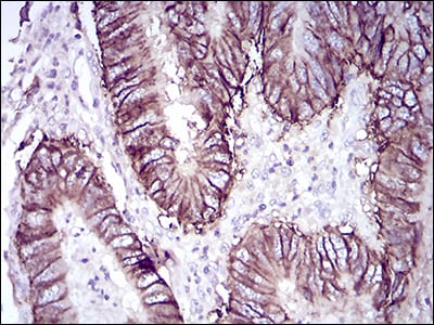
Figure 3: Immunohistochemical analysis of paraffin-embedded rectum cancer tissues using CDH17 mouse mAb with DAB staining.
Immunohistochemical analysis
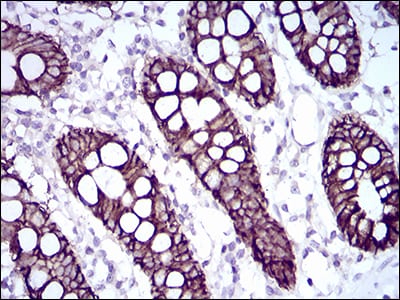
Figure 4: Immunohistochemical analysis of paraffin-embedded colon tissues using CDH17 mouse mAb with DAB staining.
Elisa
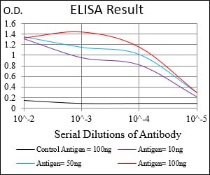
Black line: Control Antigen (100 ng); Purple line: Antigen(10ng); Blue line: Antigen (50 ng); Red line: Antigen (100 ng);
For Research Use Only. Not for use in diagnostic procedures.