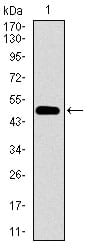
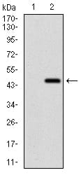
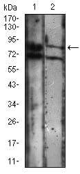
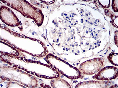
CDH16 Primary Antibody
This gene is a member of the cadherin superfamily, genes encoding calcium-dependent, membrane-associated glycoproteins. Mapped to a previously identified cluster of cadherin genes on chromosome 16q22.1, the gene localizes with superfamily members CDH1, CDH3, CDH5, CDH8 and CDH11. The protein consists of an extracellular domain containing 6 cadherin domains, a transmembrane region and a truncated cytoplasmic domain but lacks the prosequence and tripeptide HAV adhesion recognition sequence typical of most classical cadherins. Expression is exclusively in kidney, where the protein functions as the principal mediator of homotypic cellular recognition, playing a role in the morphogenic direction of tissue development. Alternatively spliced transcript variants encoding distinct isoforms have been identified.
2. Am J Physiol. 1999 Jul;277(1 Pt 2):F146-56.
![Figure 1: Western blot analysis using CDH16 mAb against human CDH16 (AA: EX(558-721)) recombinant protein. (Expected MW is 43.6 kDa)](https://cdn.shopify.com/s/files/1/0604/2661/7019/products/30659rwb_1.jpg?v=1666703224)
![Figure 2: Western blot analysis using CDH16 mAb against HEK293 (1) and CDH16 (AA: EX(558-721))-hIgGFc transfected HEK293 (2) cell lysate.](https://cdn.shopify.com/s/files/1/0604/2661/7019/products/30659twb_1.jpg?v=1666703224)
![Figure 3: Western blot analysis using CDH16 mouse mAb against A431 (1), Jurkat (2) cell lysate.](https://cdn.shopify.com/s/files/1/0604/2661/7019/products/30659lwb_1.jpg?v=1666703224)
![Figure 4: Immunofluorescence analysis of A431 cells using CDH16 mouse mAb (green). Blue: DRAQ5 fluorescent DNA dye. Red: Actin filaments have been labeled with Alexa Fluor-555 phalloidin. Secondary antibody from Fisher (Cat#: 35503)](https://cdn.shopify.com/s/files/1/0604/2661/7019/products/30659icc_1.jpg?v=1666703224)
![Figure 5: Flow cytometric analysis of A431 cells using CDH16 mouse mAb (green) and negative control (red).](https://cdn.shopify.com/s/files/1/0604/2661/7019/products/30659fcm_1.jpg?v=1666703224)
![Figure 6: Immunohistochemical analysis of paraffin-embedded kidney tissues using CDH16 mouse mAb with DAB staining.](https://cdn.shopify.com/s/files/1/0604/2661/7019/products/30659ihc_1.jpg?v=1666703224)
![Black line: Control Antigen (100 ng); Purple line: Antigen(10ng); Blue line: Antigen (50 ng); Red line: Antigen (100 ng);](https://cdn.shopify.com/s/files/1/0604/2661/7019/products/30659ea_1.jpg?v=1666703224)