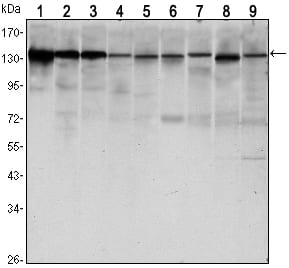
CDH1 Primary Antibody

Item Information
Catalog #
Size
Price
Description

E-Cadherin is a 120 kDa transmembrane glycoprotein that is localized in the adherens junctions of epithelial cells. There, it interacts with the cytoskeleton through the associated cytoplasmic catenin proteins. In addition to being a calcium-dependent adhesion molecule, E-Cadherin is also a critical regulator of epithelial junction formation. Its association with catenins is necessary for cell-cell adhesion. These E-cadherin/catenin complexes associate with corical actin bundles at both the zonula adherens and the lateral adhesion plaques. Tyrosine phosphorylation can disrupt these complexes, leading to changes in cell adhesion properties. E-Cadherin expression is often down-regulated in highly invasive, poorly differentiated carcinomas. Increased expression of E-Cadherin in these cells reduces invasiveness. Thus, loss of expression or function of E-Cadherin appears to be an important step in tumorigenic progression.Tissue specificity: Non-neural epithelial tissues.

Product Overview
Entrez GenelD
999
Aliases
UVO; CDHE; ECAD; LCAM; Arc-1; CD324; CDH1
Clone#
7H12
Host / Isotype
Mouse / IgG1
Species Reactivity
Human, Mouse, Monkey
Immunogen
Purified recombinant fragment of human CDH1 expressed in E. Coli.
Formulation
Ascitic fluid containing 0.03% sodium azide.
Storage
4°C; -20°C for long term storage
Product Applications
WB (Western Blot)
1/500 - 1/2000
IHC_P(Immunohistochemistry)
1/200 - 1/1000
FCM (Flow Cytometry)
1/200 - 1/400
ELISA
1/10000
References
1. Nat Genet. 2009 Dec;41(12):1330-4.
2. Zhonghua Zhong Liu Za Zhi. 2009 Jul;31(7):515-9.
3. J Biol Chem. 2010 Feb 26;285(9):6658-69.
Product Image
Western Blot
Figure 1: Western blot analysis using CDH1 mouse mAb against LNCAP (1)?A431 (2), DU145 (3), PC-3 (4), MCF-7 (5), PC-12 (6), NIH/3T3 (7), C6 (8) and COS7 (9) cell lysate.
Immunohistochemical analysis
Figure 2: Immunohistochemical analysis of paraffin-embedded gastric cancer tissues (left) and lung cancer tissues (right) using CDH1 mouse mAb with DAB staining.
Flow cytometric
Figure 3: Flow cytometric analysis of Hela cells using CDH1 mouse mAb (green) and negative control (purple).
For Research Use Only. Not for use in diagnostic procedures.