
CDC37 Primary Antibody
Item Information
Catalog #
Size
Price
Description
The protein encoded by this gene is highly similar to Cdc 37, a cell division cycle control protein of Sacchromyces cerevisiae. This protein is a molecular chaperone with specific function in cell signal transduction. It has been shown to form complex with Hsp90 and a variety of protein kinases including CDK4, CDK6, SRC, RAF-1, MOK, as well as eIF2 alpha kinases. It is thought to play a critical role in directing Hsp90 to its target kinases.
Product Overview
Entrez GenelD
11140
Aliases
P50CDC37
Clone#
6B3B7
Host / Isotype
Mouse / IgG2a
Species Reactivity
Human, Mouse
Immunogen
Purified recombinant fragment of human CDC37 (AA: 241-378) expressed in E. Coli.
Formulation
Purified antibody in PBS with 0.05% sodium azide
Storage
Store at 4°C short term. Aliquot and store at -20°C long term. Avoid freeze/thaw cycles.
Product Applications
WB (Western Blot)
1/500 - 1/2000
FCM (Flow Cytometry)
1/200 - 1/400
ELISA
1/10000
References
1.Liver Int. 2015 Apr;35(4):1403-15.
2.Oncogene. 2009 Jan 15;28(2):157-69.
2.Oncogene. 2009 Jan 15;28(2):157-69.
Product Image
Elisa

Figure 1: Black line: Control Antigen (100 ng);Purple line: Antigen (10ng); Blue line: Antigen (50 ng); Red line:Antigen (100 ng)
Western Blot

Figure 2:Western blot analysis using CDC37 mAb against human CDC37 (AA: 241-378) recombinant protein. (Expected MW is 41.6 kDa)
Western Blot

Figure 3:Western blot analysis using CDC37 mAb against HEK293 (1) and CDC37 (AA: 241-378)-hIgGFc transfected HEK293 (2) cell lysate.
Western Blot

Figure 4:Western blot analysis using CDC37 mouse mAb against K562 (1), LNcap (2), A431 (3), HEK293 (4), and C2C12 (5) cell lysate.
Flow cytometric
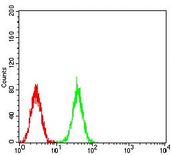
Figure 5:Flow cytometric analysis of K562 cells using CDC37 mouse mAb (green) and negative control (red).
For Research Use Only. Not for use in diagnostic procedures.