
CD89 Primary Antibody
Item Information
Catalog #
Size
Price
Description
This gene is a member of the immunoglobulin gene superfamily and encodes a receptor for the Fc region of IgA. The receptor is a transmembrane glycoprotein present on the surface of myeloid lineage cells such as neutrophils, monocytes, macrophages, and eosinophils, where it mediates immunologic responses to pathogens. It interacts with IgA-opsonized targets and triggers several immunologic defense processes, including phagocytosis, antibody-dependent cell-mediated cytotoxicity, and stimulation of the release of inflammatory mediators. Multiple alternatively spliced transcript variants encoding different isoforms have been described for this gene.
Product Overview
Entrez GenelD
2204
Aliases
FCAR; FcalphaRI; CTB-61M7.2
Clone#
2F9D8
Host / Isotype
Mouse / IgG1
Species Reactivity
Human
Immunogen
Purified recombinant fragment of human CD89 (AA: extra 22-227) expressed in E. Coli.
Formulation
Purified antibody in PBS with 0.05% sodium azide
Storage
Store at 4°C short term. Aliquot and store at -20°C long term. Avoid freeze/thaw cycles.
Product Applications
WB (Western Blot)
1/500 - 1/2000
FCM (Flow Cytometry)
1/200 - 1/400
ELISA
1/10000
References
1.Clin Exp Immunol. 2015 Sep;181(3):407-16. 2.J Immunol. 2011 Jul 15;187(2):726-32.
Product Image
Elisa
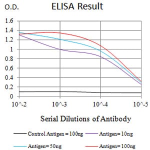
Figure 1: Black line: Control Antigen (100 ng);Purple line: Antigen (10ng); Blue line: Antigen (50 ng); Red line:Antigen (100 ng)
Western Blot
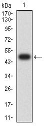
Figure 2:Western blot analysis using CD89 mAb against human CD89 (AA: extra 22-227) recombinant protein. (Expected MW is 49.4 kDa)
Western Blot
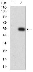
Figure 3:Western blot analysis using CD89 mAb against HEK293 (1) and CD89 (AA: extra 22-227)-hIgGFc transfected HEK293 (2) cell lysate.
Flow cytometric
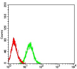
Figure 4:Flow cytometric analysis of HL-60 cells using CD89 mouse mAb (green) and negative control (red).
For Research Use Only. Not for use in diagnostic procedures.