
CD74 Primary Antibody
Item Information
Catalog #
Size
Price
Description
The protein encoded by this gene associates with class II major histocompatibility complex (MHC) and is an important chaperone that regulates antigen presentation for immune response. It also serves as cell surface receptor for the cytokine macrophage migration inhibitory factor (MIF) which, when bound to the encoded protein, initiates survival pathways and cell proliferation. This protein also interacts with amyloid precursor protein (APP) and suppresses the production of amyloid beta (Abeta). Multiple alternatively spliced transcript variants encoding different isoforms have been identified.
Product Overview
Entrez GenelD
972
Aliases
II; DHLAG; HLADG; Ia-GAMMA
Clone#
2D1B3
Host / Isotype
Mouse / IgG1
Species Reactivity
Human
Immunogen
Purified recombinant fragment of human CD74 (AA: 1-106) expressed in E. Coli.
Formulation
Purified antibody in PBS with 0.05% sodium azide
Storage
Store at 4°C short term. Aliquot and store at -20°C long term. Avoid freeze/thaw cycles.
Product Applications
WB (Western Blot)
1/500 - 1/2000
ICC (Immunocytochemistry)
1/200 - 1/1000
FCM (Flow Cytometry)
1/200 - 1/400
ELISA
1/10000
References
1. Tumour Biol. 2012 Dec;33(6):2273-7.
2. World J Gastroenterol. 2012 May 14;18(18):2253-61.
2. World J Gastroenterol. 2012 May 14;18(18):2253-61.
Product Image
Western Blot
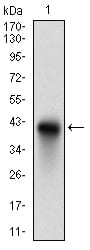
Figure 1: Western blot analysis using CD74 mAb against human CD74 recombinant protein. (Expected MW is 37.6 kDa)
Western Blot

Figure 2: Western blot analysis using CD74 mAb against HEK293 (1) and CD74 (AA: 1-106)-hIgGFc transfected HEK293 (2) cell lysate.
Immunofluorescence analysis

Figure 3: Immunofluorescence analysis of Hela cells using CD74 mouse mAb (green). Blue: DRAQ5 fluorescent DNA dye.
Western Blot

Figure 3: Western blot analysis using CD74 mouse mAb against Raji cell lysate.
Flow cytometric
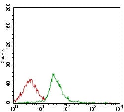
Figure 4: Flow cytometric analysis of Jurkat cells using CD74 mouse mAb (green) and negative control (red).
Elisa
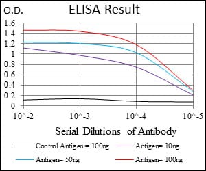
Black line: Control Antigen (100 ng); Purple line: Antigen(10ng); Blue line: Antigen (50 ng); Red line: Antigen (100 ng);
For Research Use Only. Not for use in diagnostic procedures.