

CD66A Primary Antibody
Item Information
Catalog #
Size
Price
Description
This gene encodes a member of the carcinoembryonic antigen (CEA) gene family, which belongs to the immunoglobulin superfamily. Two subgroups of the CEA family, the CEA cell adhesion molecules and the pregnancy-specific glycoproteins, are located within a 1.2 Mb cluster on the long arm of chromosome 19. Eleven pseudogenes of the CEA cell adhesion molecule subgroup are also found in the cluster. The encoded protein was originally described in bile ducts of liver as biliary glycoprotein. Subsequently, it was found to be a cell-cell adhesion molecule detected on leukocytes, epithelia, and endothelia. The encoded protein mediates cell adhesion via homophilic as well as heterophilic binding to other proteins of the subgroup. Multiple cellular activities have been attributed to the encoded protein, including roles in the differentiation and arrangement of tissue three-dimensional structure, angiogenesis, apoptosis, tumor suppression, metastasis, and the modulation of innate and adaptive immune responses. Multiple transcript variants encoding different isoforms have been reported, but the full-length nature of all variants has not been defined.
Product Overview
Entrez GenelD
634
Aliases
CEACAM1; BGP; BGP1; BGPI
Clone#
7B9G3
Host / Isotype
Mouse / IgG2b
Species Reactivity
Human
Immunogen
Purified recombinant fragment of human CD66A (AA: extra 65-201) expressed in E. Coli.
Formulation
Purified antibody in PBS with 0.05% sodium azide
Storage
Store at 4°C short term. Aliquot and store at -20°C long term. Avoid freeze/thaw cycles.
Product Applications
WB (Western Blot)
1/500 - 1/2000
IHC_P(Immunohistochemistry)
1/200 - 1/1000
FCM (Flow Cytometry)
1/200 - 1/400
ELISA
1/10000
References
1.J Biol Chem. 2016 Sep 16;291(38):20085-95.
2.PLoS One. 2016 Apr 13;11(4):e0153601.
2.PLoS One. 2016 Apr 13;11(4):e0153601.
Product Image
Elisa

Figure 1: Black line: Control Antigen (100 ng);Purple line: Antigen (10ng); Blue line: Antigen (50 ng); Red line:Antigen (100 ng)
Western Blot

Figure 2:Western blot analysis using CD66A mAb against human CD66A (AA: extra 65-201) recombinant protein. (Expected MW is 41.2 kDa)
Western Blot

Figure 3:Western blot analysis using CD66A mAb against HEK293 (1) and CD66A (AA: extra 65-201)-hIgGFc transfected HEK293 (2) cell lysate.
Western Blot

Figure 4:Western blot analysis using CD66A mouse mAb against HepG2 (1), SK-MES-1 (2), Hela (3), and A549 (4) cell lysate.
Flow cytometric
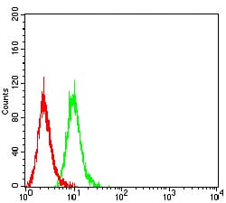
Figure 5:Flow cytometric analysis of Hela cells using CD66A mouse mAb (green) and negative control (red).
Immunohistochemical analysis

Figure 6:Immunohistochemical analysis of paraffin-embedded lung cancer tissues using CD66A mouse mAb with DAB staining.
Immunohistochemical analysis

Figure 7:Immunohistochemical analysis of paraffin-embedded cervical cancer tissues using CD66A mouse mAb with DAB staining.
For Research Use Only. Not for use in diagnostic procedures.