

CD64 Primary Antibody
Item Information
Catalog #
Size
Price
Description
This gene encodes a protein that plays an important role in the immune response. This protein is a high-affinity Fc-gamma receptor. The gene is one of three related gene family members located on chromosome 1.
Product Overview
Entrez GenelD
2209
Aliases
FCGR1A; FCRI; CD64A; IGFR1
Clone#
5F5F8
Host / Isotype
Mouse / IgG1
Species Reactivity
Human
Immunogen
Purified recombinant fragment of human CD64 (AA: extra 16-145) expressed in E. Coli.
Formulation
Purified antibody in PBS with 0.05% sodium azide
Storage
Store at 4°C short term. Aliquot and store at -20°C long term. Avoid freeze/thaw cycles.
Product Applications
WB (Western Blot)
1/500 - 1/2000
IHC_P(Immunohistochemistry)
1/200 - 1/1000
FCM (Flow Cytometry)
1/200 - 1/400
ELISA
1/10000
References
1.Int J Lab Hematol. 2016 Oct;38(5):576-84. 2.Inflamm Res. 2016 Jul;65(7):579-85.
Product Image
Elisa
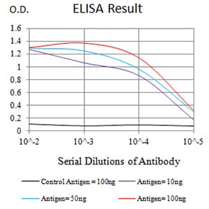
Figure 1: Black line: Control Antigen (100 ng);Purple line: Antigen (10ng); Blue line: Antigen (50 ng); Red line:Antigen (100 ng)
Western Blot
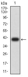
Figure 2:Western blot analysis using CD64 mAb against human CD64 (AA: extra 16-145) recombinant protein. (Expected MW is 40.6 kDa)
Western Blot
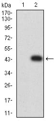
Figure 3:Western blot analysis using CD64 mAb against HEK293 (1) and CD64 (AA: extra 16-145)-hIgGFc transfected HEK293 (2) cell lysate.
Flow cytometric
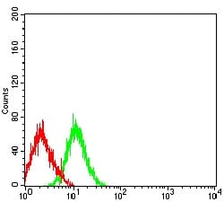
Figure 4:Flow cytometric analysis of HL-60 cells using CD64 mouse mAb (green) and negative control (red).
Immunohistochemical analysis
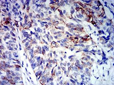
Figure 5:Immunohistochemical analysis of paraffin-embedded bladder cancer tissues using CD64 mouse mAb with DAB staining.
For Research Use Only. Not for use in diagnostic procedures.