

CD6 Primary Antibody
Item Information
Catalog #
Size
Price
Description
This gene encodes a protein found on the outer membrane of T-lymphocytes as well as some other immune cells. The encoded protein contains three scavenger receptor cysteine-rich (SRCR) domains and a binding site for an activated leukocyte cell adhesion molecule. The gene product is important for continuation of T cell activation. This gene may be associated with susceptibility to multiple sclerosis (PMID: 19525953, 21849685). Multiple transcript variants encoding different isoforms have been found for this gene.
Product Overview
Entrez GenelD
923
Aliases
TP120; FLJ44171
Clone#
6B9
Host / Isotype
Mouse / IgG1
Species Reactivity
Human
Immunogen
Purified recombinant fragment of human CD6 (AA: 472-668) expressed in E. Coli.
Formulation
Purified antibody in PBS with 0.05% sodium azide
Storage
Store at 4°C short term. Aliquot and store at -20°C long term. Avoid freeze/thaw cycles.
Product Applications
WB (Western Blot)
1/500 - 1/2000
IHC_P(Immunohistochemistry)
1/200 - 1/1000
ELISA
1/10000
References
1.J Autoimmun. 2010 Dec;35(4):336-41. 2.Proc Natl Acad Sci U S A. 2007 Jul 10;104(28):11724-9.
Product Image
Western Blot

Figure 1: Western blot analysis using CD6 mAb against human CD6 recombinant protein. (Expected MW is 46.9 kDa)
Western Blot
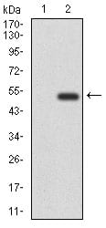
Figure 2: Western blot analysis using CD6 mAb against HEK293 (1) and CD6 (AA: 472-668)-hIgGFc transfected HEK293 (2) cell lysate.
Immunohistochemical analysis
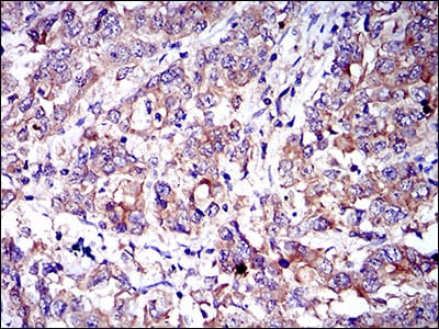
Figure 3: Immunohistochemical analysis of paraffin-embedded stomach cancer tissues using CD6 mouse mAb with DAB staining.
Elisa

Black line: Control Antigen (100 ng); Purple line: Antigen(10ng); Blue line: Antigen (50 ng); Red line: Antigen (100 ng);
For Research Use Only. Not for use in diagnostic procedures.