

CD57 Primary Antibody
Item Information
Catalog #
Size
Price
Description
The protein encoded by this gene is a member of the glucuronyltransferase gene family. These enzymes exhibit strict acceptor specificity, recognizing nonreducing terminal sugars and their anomeric linkages. This gene product functions as the key enzyme in a glucuronyl transfer reaction during the biosynthesis of the carbohydrate epitope HNK-1 (human natural killer-1, also known as CD57 and LEU7). Alternate transcriptional splice variants have been characterized. [provided by RefSeq, Jul 2008]
Product Overview
Entrez GenelD
27087
Aliases
NK1; CD57; HNK1; LEU7; NK-1; GLCATP; GLCUATP
Clone#
1A8B9
Host / Isotype
Mouse / Mouse IgG2b
Immunogen
Purified recombinant fragment of human CD57 (AA: 28-334) expressed in E. Coli.
Formulation
Purified antibody in PBS with 0.05% sodium azide
Storage
Store at 4°C short term. Aliquot and store at -20°C long term. Avoid freeze/thaw cycles.
Product Applications
WB (Western Blot)
1/500 - 1/2000
IHC_P(Immunohistochemistry)
1/200-1/1000
FCM (Flow Cytometry)
1/200-1/400
ELISA
1/10000
References
1,Mol Neurobiol. 2019 Sep;56(9):6581-6585. 2,J Acquir Immune Defic Syndr. 2016 Jun 1;72(2):133-7.
Product Image
ELISA
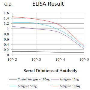
Figure 1: Black line: Control Antigen (100 ng);Purple line: Antigen (10ng); Blue line: Antigen (50 ng); Red line: Antigen (100 ng)
WESTERN BLOT
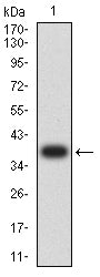
Figure 2: Western blot analysis using CD57 mAb against human CD57 (AA: 28-334) recombinant protein. (Expected MW is 37.9kDa)
WESTERN BLOT
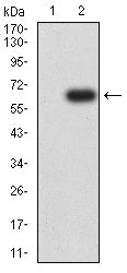
Figure 3: Western blot analysis using CD57 mAb against HEK293-6e (1) and CD57 (AA: 28-334)-hIgGFc transfected HEK293-6e (2) cell lysate.
FLOW CYTOMETRY
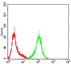
Figure 4: Flow cytometric analysis of Hela cells using CD57 mouse mAb (green) and negative control (red).
IMMUNOHISTOCHEMISTRY
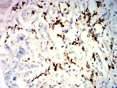
Figure 5: Immunohistochemical analysis of paraffin-embedded rectal cancer tissues using CD57 mouse mAb with DAB staining.
IMMUNOHISTOCHEMISTRY

Figure 6: Immunohistochemical analysis of paraffin-embedded brain tissues using CD57 mouse mAb with DAB staining.
For Research Use Only. Not for use in diagnostic procedures.