

CD54 Primary Antibody
Item Information
Catalog #
Size
Price
Description
This gene encodes a cell surface glycoprotein which is typically expressed on endothelial cells and cells of the immune system. It binds to integrins of type CD11a / CD18, or CD11b / CD18 and is also exploited by Rhinovirus as a receptor.
Product Overview
Entrez GenelD
3383
Aliases
BB2; CD54; P3.58
Clone#
1D5D5
Host / Isotype
Mouse / Mouse IgG2b
Species Reactivity
Human
Immunogen
Purified recombinant fragment of human CD54 (AA: 28-163) expressed in E. Coli.
Formulation
Purified antibody in PBS with 0.05% sodium azide
Storage
Store at 4°C short term. Aliquot and store at -20°C long term. Avoid freeze/thaw cycles.
Product Applications
WB (Western Blot)
1/500 - 1/2000
IHC_P(Immunohistochemistry)
1/200 - 1/1000
FCM (Flow Cytometry)
1/200 - 1/400
ELISA
1/10000
References
1.Dev Cell. 2022 Feb 7;57(3):329-343.e7.
2.Korean J Gastroenterol. 2022 Apr 25;79(4):170-176.
2.Korean J Gastroenterol. 2022 Apr 25;79(4):170-176.
Product Image
Elisa

Figure 1:Black line: Control Antigen (100 ng);Purple line: Antigen (10ng); Blue line: Antigen (50 ng); Red line:Antigen (100 ng)
Western Blot

Figure 2:Western blot analysis using CD54 mAb against human CD54 (AA: 28-163) recombinant protein. (Expected MW is 40.8 kDa)
Western Blot

Figure 3:Western blot analysis using CD54 mAb against HEK293-6e (1) and CD54 (AA: 28-163)-hIgGFc transfected HEK293-6e (2) cell lysate.
Western Blot

Figure 4:Western blot analysis using CD54 mouse mAb against Raji (1) cell lysate.
Immunofluorescence analysis

Figure 5:Flow cytometric analysis of Jurkat cells using CD54 mouse mAb (green) and negative control (red).
Immunohistochemical analysis

Figure 6:Immunohistochemical analysis of paraffin-embedded liver cancer tissues using CD54 mouse mAb with DAB staining.
Immunohistochemical analysis
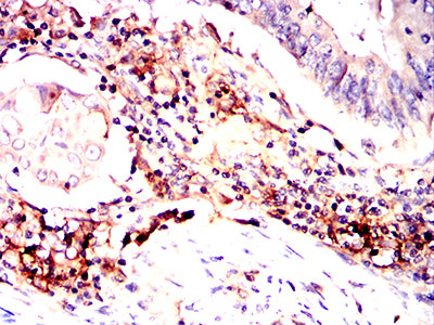
Figure 7:Immunohistochemical analysis of paraffin-embedded rectal cancer tissues using CD54 mouse mAb with DAB staining.
Immunohistochemical analysis

Figure 8:Immunohistochemical analysis of paraffin-embedded renal tissue tissues using CD54 mouse mAb with DAB staining.
Immunohistochemical analysis

Figure 9:Immunohistochemical analysis of paraffin-embedded esophageal tissue tissues using CD54 mouse mAb with DAB staining.
Immunofluorescence analysis

Figure 10:Flow cytometric analysis of Raji cells using CD54 mouse mAb (green) and negative control (red).
For Research Use Only. Not for use in diagnostic procedures.