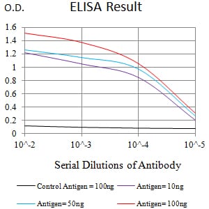
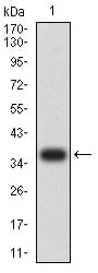
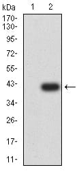
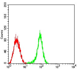
CD53 Primary Antibody

Item Information
Catalog #
Size
Price
Description

The protein encoded by this gene is a member of the transmembrane 4 superfamily, also known as the tetraspanin family. Most of these members are cell-surface proteins that are characterized by the presence of four hydrophobic domains. The proteins mediate signal transduction events that play a role in the regulation of cell development, activation, growth and motility. This encoded protein is a cell surface glycoprotein that is known to complex with integrins. It contributes to the transduction of CD2-generated signals in T cells and natural killer cells and has been suggested to play a role in growth regulation. Familial deficiency of this gene has been linked to an immunodeficiency associated with recurrent infectious diseases caused by bacteria, fungi and viruses. Alternative splicing results in multiple transcript variants.

Product Overview
Entrez GenelD
963
Aliases
MOX44; TSPAN25
Clone#
1H2C7
Host / Isotype
Mouse / IgG1
Species Reactivity
Human
Immunogen
Purified recombinant fragment of human CD53 (AA: extra mix) expressed in E. Coli.
Formulation
Purified antibody in PBS with 0.05% sodium azide
Storage
4°C; -20°C for long term storage
Product Applications
WB (Western Blot)
1/500 - 1/2000
IHC_P(Immunohistochemistry)
1/200 - 1/1000
FCM (Flow Cytometry)
1/200 - 1/400
ELISA
1/10000
References
1.Biochim Biophys Acta. 2013 Apr;1830(4):3011-8.
2.Eur J Hum Genet. 2010 Aug;18(8):953-9.
Product Image
Elisa
Figure 1:Black line: Control Antigen (100 ng);Purple line: Antigen (10ng); Blue line: Antigen (50 ng); Red line:Antigen (100 ng)
Western Blot
Figure 2:Western blot analysis using CD53 mAb against human CD53 (AA: extra mix) recombinant protein. (Expected MW is 37 kDa)
Western Blot
Figure 3:Western blot analysis using CD53 mAb against HEK293 (1) and CD53 (AA: extra mix)-hIgGFc transfected HEK293 (2) cell lysate.
Flow cytometric
Figure 4:Flow cytometric analysis of HL-60 cells using CD53 mouse mAb (green) and negative control (red).
Immunohistochemical analysis
Figure 5:Immunohistochemical analysis of paraffin-embedded rectum cancer tissues using CD53 mouse mAb with DAB staining.
For Research Use Only. Not for use in diagnostic procedures.