

CD50 Primary Antibody
Item Information
Catalog #
Size
Price
Description
The protein encoded by this gene is a member of the intercellular adhesion molecule (ICAM) family. All ICAM proteins are type I transmembrane glycoproteins, contain 2-9 immunoglobulin-like C2-type domains, and bind to the leukocyte adhesion LFA-1 protein. This protein is constitutively and abundantly expressed by all leucocytes and may be the most important ligand for LFA-1 in the initiation of the immune response. It functions not only as an adhesion molecule, but also as a potent signalling molecule. Alternative splicing results in multiple transcript variants encoding different isoforms.
Product Overview
Entrez GenelD
3385
Aliases
ICAM3; CDW50; ICAM-R
Clone#
2D11D1
Host / Isotype
Mouse / IgG1
Species Reactivity
Human
Immunogen
Purified recombinant fragment of human CD50 (AA: extra 30-203) expressed in E. Coli.
Formulation
Purified antibody in PBS with 0.05% sodium azide
Storage
Store at 4°C short term. Aliquot and store at -20°C long term. Avoid freeze/thaw cycles.
Product Applications
WB (Western Blot)
1/500 - 1/2000
FCM (Flow Cytometry)
1/200 - 1/400
ELISA
1/10000
References
1.Biochem Biophys Res Commun. 2013 Nov 15;441(2):507-13.
2.Apoptosis. 2013 Oct;18(10):1235-51.
2.Apoptosis. 2013 Oct;18(10):1235-51.
Product Image
Elisa

Figure 1: Black line: Control Antigen (100 ng);Purple line: Antigen (10ng); Blue line: Antigen (50 ng); Red line:Antigen (100 ng)
Western Blot

Figure 2:Western blot analysis using CD50 mAb against human CD50 (AA: extra 30-203) recombinant protein. (Expected MW is 44.8 kDa)
Western Blot
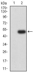
Figure 3:Western blot analysis using CD50 mAb against HEK293 (1) and CD50 (AA: extra 30-203)-hIgGFc transfected HEK293 (2) cell lysate.
Flow cytometric
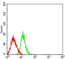
Figure 4:Flow cytometric analysis of HL-60 cells using CD50 mouse mAb (green) and negative control (red).
For Research Use Only. Not for use in diagnostic procedures.