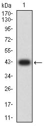
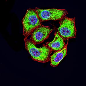
CD46Primary Antibody

Item Information
Catalog #
Size
Price
Description

The protein encoded by this gene is a type I membrane protein and is a regulatory part of the complement system. The encoded protein has cofactor activity for inactivation of complement components C3b and C4b by serum factor I, which protects the host cell from damage by complement. In addition, the encoded protein can act as a receptor for the Edmonston strain of measles virus, human herpesvirus-6, and type IV pili of pathogenic Neisseria. Finally, the protein encoded by this gene may be involved in the fusion of the spermatozoa with the oocyte during fertilization. Mutations at this locus have been associated with susceptibility to hemolytic uremic syndrome. Alternatively spliced transcript variants encoding different isoforms have been described.

Product Overview
Entrez GenelD
4179
Aliases
MCP; TLX; AHUS2; MIC10; TRA2.10
Clone#
5A8C11
Host / Isotype
Mouse / IgG1
Species Reactivity
Human
Immunogen
Purified recombinant fragment of human CD46 (AA: extra 35-179) expressed in E. Coli.
Formulation
Purified antibody in PBS with 0.05% sodium azide
Storage
4°C; -20°C for long term storage
Product Applications
WB (Western Blot)
1/500 - 1/2000
ICC (Immunocytochemistry)
1/100 - 1/500
FCM (Flow Cytometry)
1/200 - 1/400
ELISA
1/10000
References
1.J Clin Invest. 2016 Dec 1;126(12):4640-4653.
2.PLoS One. 2012;7(10):e48486.
Product Image
Elisa
Figure 1:Black line: Control Antigen (100 ng);Purple line: Antigen (10ng); Blue line: Antigen (50 ng); Red line:Antigen (100 ng)
Western Blot
Figure 2:Western blot analysis using CD46 mAb against human CD46 (AA: extra 35-179) recombinant protein. (Expected MW is 42.4 kDa)
Western Blot
Figure 3:Western blot analysis using CD46 mAb against HEK293 (1) and CD46 (AA: extra 35-179)-hIgGFc transfected HEK293 (2) cell lysate.
Western Blot
Figure 4:Western blot analysis using CD46 mouse mAb against SW480 (1) and MCF-7 (2) cell lysate.
Immunofluorescence analysis
Figure 5:Immunofluorescence analysis of Hela cells using CD46 mouse mAb (green). Blue: DRAQ5 fluorescent DNA dye. Red: Actin filaments have been labeled with Alexa Fluor- 555 phalloidin. Secondary antibody from Fisher (Cat#: 35503)
Flow cytometric
Figure 6:Flow cytometric analysis of HL-60 cells using CD46 mouse mAb (green) and negative control (red).
For Research Use Only. Not for use in diagnostic procedures.