

CD44 Primary Antibody
Item Information
Catalog #
Size
Price
Description
The protein encoded by this gene is a cell-surface glycoprotein involved in cell-cell interactions, cell adhesion and migration. It is a receptor for hyaluronic acid (HA) and can also interact with other ligands, such as osteopontin, collagens, and matrix metalloproteinases (MMPs). This protein participates in a wide variety of cellular functions including lymphocyte activation, recirculation and homing, hematopoiesis, and tumor metastasis. Transcripts for this gene undergo complex alternative splicing that results in many functionally distinct isoforms, however, the full length nature of some of these variants has not been determined. Alternative splicing is the basis for the structural and functional diversity of this protein, and may be related to tumor metastasis.
Product Overview
Entrez GenelD
960
Aliases
IN; LHR; MC56; MDU2; MDU3; MIC4; Pgp1; CDW44; CSPG8; HCELL; HUTCH-I; ECMR-III
Clone#
7F4F1
Host / Isotype
Mouse / IgG1
Species Reactivity
Human
Immunogen
Purified recombinant fragment of human CD44 (AA: extra 36-194) expressed in E. Coli.
Formulation
Purified antibody in PBS with 0.05% sodium azide
Storage
Store at 4°C short term. Aliquot and store at -20°C long term. Avoid freeze/thaw cycles.
Product Applications
WB (Western Blot)
1/500 - 1/2000
IHC_P(Immunohistochemistry)
1/200 - 1/1000
FCM (Flow Cytometry)
1/200 - 1/400
ELISA
1/10000
References
1.Mol Med Rep. 2016 Oct;14(4):3159-67.
2.Mol Cancer Res. 2016 Apr;14(4):344-53.
2.Mol Cancer Res. 2016 Apr;14(4):344-53.
Product Image
Elisa

Figure 1: Black line: Control Antigen (100 ng);Purple line: Antigen (10ng); Blue line: Antigen (50 ng); Red line:Antigen (100 ng)
Western Blot

Figure 4:Western blot analysis using CD44 mouse mAb against Hela (1), PANC-1 (2), HUVEC (3), and HUVE-12 (4) cell lysate.
Flow cytometric

Figure 5:Flow cytometric analysis of Hela cells using CD44 mouse mAb (green) and negative control (red).
Immunohistochemical analysis

Figure 6:Immunohistochemical analysis of paraffin-embedded bladder cancer tissues using CD44 mouse mAb with DAB staining.
Western Blot

Figure 6:Western blot analysis using CD44 mAb against human CD44 (AA: extra 36-194) recombinant protein. (Expected MW is 43.3 kDa)
Immunohistochemical analysis

Figure 7:Immunohistochemical analysis of paraffin-embedded rectum cancer tissues using CD44 mouse mAb with DAB staining.
Western Blot
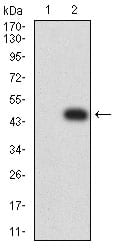
Figure 7:Western blot analysis using CD44 mAb against HEK293 (1) and CD44 (AA: extra 36-194)-hIgGFc transfected HEK293 (2) cell lysate.
For Research Use Only. Not for use in diagnostic procedures.