

CD42A Primary Antibody
Item Information
Catalog #
Size
Price
Description
This gene encodes a small membrane glycoprotein found on the surface of human platelets. It forms a 1-to-1 noncovalent complex with glycoprotein Ib, a platelet surface membrane glycoprotein complex that functions as a receptor for von Willebrand factor. The complete receptor complex includes noncovalent association of the alpha and beta subunits with the protein encoded by this gene and platelet glycoprotein V. Defects in this gene are a cause of Bernard-Soulier syndrome, also known as giant platelet disease. These patients have unusually large platelets and have a clinical bleeding tendency.
Product Overview
Entrez GenelD
2815
Aliases
GP9; GPIX
Clone#
4E2G7
Species Reactivity
Human
Immunogen
Purified recombinant fragment of human CD42A (AA: extra 17-147) expressed in E. Coli.
Formulation
Purified antibody in PBS with 0.05% sodium azide
Storage
Store at 4°C short term. Aliquot and store at -20°C long term. Avoid freeze/thaw cycles.
Product Applications
WB (Western Blot)
1/500 - 1/2000
FCM (Flow Cytometry)
1/200 - 1/400
ELISA
1/10000
References
1.J Biol Chem. 2015 Sep 4;290(36):22155-62.
2.Blood. 2011 Nov 10;118(19):5292-301.
2.Blood. 2011 Nov 10;118(19):5292-301.
Product Image
Elisa
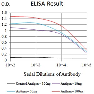
Figure 1:Black line: Control Antigen (100 ng);Purple line: Antigen (10ng); Blue line: Antigen (50 ng); Red line:Antigen (100 ng)
Western Blot

Figure 2:Western blot analysis using CD42A mAb against human CD42A (AA: extra 17-147) recombinant protein. (Expected MW is 40.3 kDa)
Western Blot

Figure 3:Western blot analysis using CD42A mAb against HEK293 (1) and CD42A (AA: extra 17-147)-hIgGFc transfected HEK293 (2) cell lysate.
Flow cytometric

Figure 4:Flow cytometric analysis of HL-60 cells using CD42A mouse mAb (green) and negative control (red).
For Research Use Only. Not for use in diagnostic procedures.