CD40 Primary Antibody

Item Information
Catalog #
Size
Price
Description

This gene is a member of the TNF-receptor superfamily. The encoded protein is a receptor on antigen-presenting cells of the immune system and is essential for mediating a broad variety of immune and inflammatory responses including T cell-dependent immunoglobulin class switching, memory B cell development, and germinal center formation. AT-hook transcription factor AKNA is reported to coordinately regulate the expression of this receptor and its ligand, which may be important for homotypic cell interactions. Adaptor protein TNFR2 interacts with this receptor and serves as a mediator of the signal transduction. The interaction of this receptor and its ligand is found to be necessary for amyloid-beta-induced microglial activation, and thus is thought to be an early event in Alzheimer disease pathogenesis. Mutations affecting this gene are the cause of autosomal recessive hyper-IgM immunodeficiency type 3 (HIGM3). Multiple alternatively spliced transcript variants of this gene encoding distinct isoforms have been reported. [provided by RefSeq, Nov 2014]
Product Overview
Entrez GenelD
958
Aliases
p50; Bp50; CDW40; TNFRSF5
Clone#
8C8A5
Host / Isotype
Mouse / Mouse IgG1
Immunogen
Purified recombinant fragment of human CD40 (AA: extra(21-193)) expressed in E. Coli.
Formulation
Purified antibody in PBS with 0.05% sodium azide
Storage
4°C; -20°C for long term storage
Product Applications
WB (Western Blot)
1/500 - 1/2000
FCM (Flow Cytometry)
1/200-1/400
ELISA
1/10000
References
1,Life Sci. 2020 Aug 1;254:117774. 2,Mol Genet Genomic Med. 2019 Dec;7(12):e1014.
Product Image
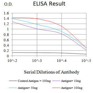
ELISA
Figure 1: Black line: Control Antigen (100 ng);Purple line: Antigen (10ng); Blue line: Antigen (50 ng); Red line: Antigen (100 ng)
WESTERN BLOT
Figure 2: Western blot analysis using CD40 mAb against human CD40 (AA: extra(21-193)) recombinant protein. (Expected MW is 22 kDa)
WESTERN BLOT
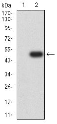
Figure 3: Western blot analysis using CD40 mAb against HEK293-6e (1) and CD40 (AA: extra(21-193))-hIgGFc transfected HEK293-6e (2) cell lysate.
WESTERN BLOT
Figure 4: Western blot analysis using CD40 mouse mAb against Raji cell lysate.
FLOW CYTOMETRY
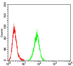
Figure 5: Flow cytometric analysis of Raji cells using CD40 mouse mAb (green) and negative control (red).
For Research Use Only. Not for use in diagnostic procedures.