
CD344 Primary Antibody
Item Information
Catalog #
Size
Price
Description
This gene is a member of the frizzled gene family. Members of this family encode seven-transmembrane domain proteins that are receptors for the Wingless type MMTV integration site family of signaling proteins. Most frizzled receptors are coupled to the beta-catenin canonical signaling pathway. This protein may play a role as a positive regulator of the Wingless type MMTV integration site signaling pathway. A transcript variant retaining intronic sequence and encoding a shorter isoform has been described, however, its expression is not supported by other experimental evidence.
Product Overview
Entrez GenelD
8322
Aliases
FZD4; Fz4; EVR1; FEVR; Fz-4; FzE4; GPCR; hFz4; FZD4S
Clone#
2F3A7
Host / Isotype
Mouse / IgG1
Species Reactivity
Human
Immunogen
Purified recombinant fragment of human CD344 (AA: extra 37-222) expressed in E. Coli.
Formulation
Purified antibody in PBS with 0.05% sodium azide
Storage
Store at 4°C short term. Aliquot and store at -20°C long term. Avoid freeze/thaw cycles.
Product Applications
WB (Western Blot)
1/500 - 1/2000
FCM (Flow Cytometry)
1/200 - 1/400
ELISA
1/10000
References
1.Ophthalmic Genet. 2017 Jul-Aug;38(4):380-382.
2.Mol Pharmacol. 2016 Oct;90(4):447-59.
2.Mol Pharmacol. 2016 Oct;90(4):447-59.
Product Image
Elisa

Figure 1:Black line: Control Antigen (100 ng);Purple line: Antigen (10ng); Blue line: Antigen (50 ng); Red line:Antigen (100 ng)
Western Blot

Figure 2:Western blot analysis using CD344 mAb against human CD344 (AA: extra 37-222) recombinant protein. (Expected MW is 47 kDa)
Western Blot

Figure 3:Western blot analysis using CD344 mAb against HEK293 (1) and CD344 (AA: extra 37-222)-hIgGFc transfected HEK293 (2) cell lysate.
Flow cytometric
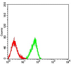
Figure 4:Flow cytometric analysis of HL-60 cells using CD344 mouse mAb (green) and negative control (red).
For Research Use Only. Not for use in diagnostic procedures.