
CD328 Primary Antibody
Item Information
Catalog #
Size
Price
Description
SIGLEC7 (Sialic Acid Binding Ig Like Lectin 7) is a Protein Coding gene. Diseases associated with SIGLEC7 include Congenital Disorder Of Glycosylation, Type Iic. Among its related pathways are Hematopoietic Stem Cells and Lineage-specific Markers and Innate Immune System. Gene Ontology (GO) annotations related to this gene include carbohydrate binding. An important paralog of this gene is SIGLEC12.
Product Overview
Entrez GenelD
27036
Aliases
p75; QA79; AIRM1; CD328; AIRM-1; CDw328; D-siglec; SIGLEC-7; SIGLECP2; SIGLEC19P; p75/AIRM1
Clone#
6G8B10
Host / Isotype
Mouse / Mouse IgG1
Immunogen
Purified recombinant fragment of human CD328 (AA: extra(19-142)) expressed in E. Coli.
Formulation
Purified antibody in PBS with 0.05% sodium azide
Storage
Store at 4°C short term. Aliquot and store at -20°C long term. Avoid freeze/thaw cycles.
Product Applications
WB (Western Blot)
1/500 - 1/2000
IHC_P(Immunohistochemistry)
1/200-1/1000
FCM (Flow Cytometry)
1/200-1/400
ELISA
1/10000
References
1,Allergy. 2019 Jul;74(7):1257-1265. 2,Int J Mol Sci. 2018 Apr 4;19(4):1073.
Product Image
ELISA
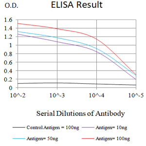
Figure 1: Black line: Control Antigen (100 ng);Purple line: Antigen (10ng); Blue line: Antigen (50 ng); Red line: Antigen (100 ng)
WESTERN BLOT
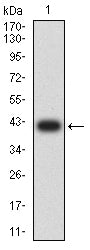
Figure 2: Western blot analysis using CD328 mAb against human CD328 (AA: extra(19-142)) recombinant protein. (Expected MW is 40.2 kDa)
WESTERN BLOT
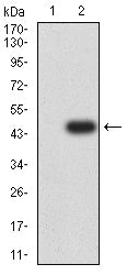
Figure 3: Western blot analysis using CD328 mAb against HEK293-6e (1) and CD328 (AA: extra(19-142))-hIgGFc transfected HEK293-6e (2) cell lysate.
FLOW CYTOMETRY
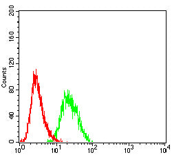
Figure 4: Flow cytometric analysis of HL-60 cells using CD328 mouse mAb (green) and negative control (red).
IMMUNOHISTOCHEMISTRY
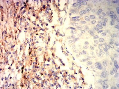
Figure 5: Immunohistochemical analysis of paraffin-embedded lung cancer tissues using CD328 mouse mAb with DAB staining.
For Research Use Only. Not for use in diagnostic procedures.