

CD327 Primary Antibody
Item Information
Catalog #
Size
Price
Description
This gene encodes a member of the SIGLEC (sialic acid binding immunoglobulin-like lectin) family of proteins. The encoded transmembrane receptor binds sialyl-TN glycans and leptin. Placental expression of the encoded protein is upregulated in preeclampsia.
Product Overview
Entrez GenelD
946
Aliases
SIGLEC6; CD33L; OBBP1; CD33L1; CD33L2; CDW327
Clone#
3C11C12G1
Host / Isotype
Mouse / IgG2b
Species Reactivity
Human
Immunogen
Purified recombinant fragment of human CD327 (AA: extra 27-347) expressed in E. Coli.
Formulation
Purified antibody in PBS with 0.05% sodium azide
Storage
Store at 4°C short term. Aliquot and store at -20°C long term. Avoid freeze/thaw cycles.
Product Applications
WB (Western Blot)
1/500 - 1/2000
IHC_P(Immunohistochemistry)
1/200 - 1/1000
FCM (Flow Cytometry)
1/200 - 1/400
ELISA
1/10000
References
1.Reprod Sci. 2013 Jun;20(6):646-53.
2.Endocr Relat Cancer. 2012 Nov 19;19(6):827-40.
2.Endocr Relat Cancer. 2012 Nov 19;19(6):827-40.
Product Image
Elisa

Figure 1:Black line: Control Antigen (100 ng);Purple line: Antigen (10ng); Blue line: Antigen (50 ng); Red line:Antigen (100 ng)
Western Blot

Figure 2:Western blot analysis using CD327 mAb against human CD327 (AA: extra 27-347) recombinant protein. (Expected MW is 61.4 kDa)
Western Blot

Figure 3:Western blot analysis using CD327 mAb against HEK293 (1) and CD327 (AA: extra 27-347)-hIgGFc transfected HEK293 (2) cell lysate.
Flow cytometric

Figure 4:Flow cytometric analysis of HL-60 cells using CD327 mouse mAb (green) and negative control (red).
Immunohistochemical analysis
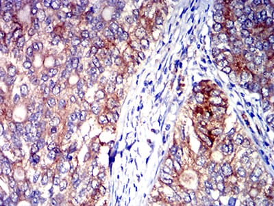
Figure 5:Immunohistochemical analysis of paraffin-embedded cervical cancer tissues using CD327 mouse mAb with DAB staining.
Immunohistochemical analysis

Figure 6:Immunohistochemical analysis of paraffin-embedded rectum cancer tissues using CD327 mouse mAb with DAB staining.
For Research Use Only. Not for use in diagnostic procedures.