

CD322 Primary Antibody
Item Information
Catalog #
Size
Price
Description
This gene belongs to the immunoglobulin superfamily, and the junctional adhesion molecule (JAM) family. The protein encoded by this gene is a type I membrane protein that is localized at the tight junctions of both epithelial and endothelial cells. It acts as an adhesive ligand for interacting with a variety of immune cell types, and may play a role in lymphocyte homing to secondary lymphoid organs. Alternatively spliced transcript variants have been found for this gene.
Product Overview
Entrez GenelD
58494
Aliases
JAM2; JAMB; JAM-B; VEJAM; PRO245; VE-JAM; C21orf43
Clone#
7E4C5
Host / Isotype
Mouse / IgG1
Species Reactivity
Human
Immunogen
Purified recombinant fragment of human CD322 (AA: extra 29-238) expressed in E. Coli.
Formulation
Purified antibody in PBS with 0.05% sodium azide
Storage
Store at 4°C short term. Aliquot and store at -20°C long term. Avoid freeze/thaw cycles.
Product Applications
WB (Western Blot)
1/500 - 1/2000
ICC (Immunocytochemistry)
1/100
FCM (Flow Cytometry)
1/200 - 1/400
ELISA
1/10000
References
1.Oncol Rep. 2016 Jul;36(1):3-9.
2.Int J Oncol. 2016 Mar;48(3):929-36.
2.Int J Oncol. 2016 Mar;48(3):929-36.
Product Image
Elisa
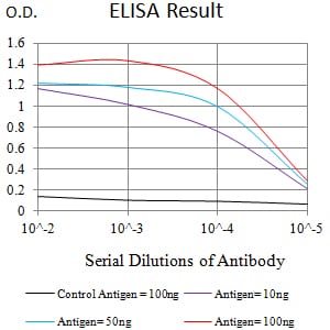
Figure 1:Black line: Control Antigen (100 ng);Purple line: Antigen (10ng); Blue line: Antigen (50 ng); Red line:Antigen (100 ng)
Western Blot

Figure 2:Western blot analysis using CD322 mAb against human CD322 (AA: extra 29-238) recombinant protein. (Expected MW is 53.3 kDa)
Western Blot

Figure 3:Western blot analysis using CD322 mAb against HEK293 (1) and CD322 (AA: extra 29-238)-hIgGFc transfected HEK293 (2) cell lysate.
Western Blot

Figure 4:Western blot analysis using CD322 mouse mAb against NIH/3T3 (1), Ramos (2), and HepG2 (3) cell lysate.
Flow cytometric

Figure 5:Flow cytometric analysis of HL-60 cells using CD322 mouse mAb (green) and negative control (red).
Immunofluorescence analysis
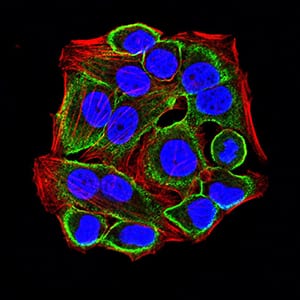
Figure 6:Immunofluorescence analysis of Hela cells using CD322 mouse mAb (green). Blue: DRAQ5 fluorescent DNA dye. Red: Actin filaments have been labeled with Alexa Fluor- 555 phalloidin. Secondary antibody from Fisher (Cat#: 35503)
For Research Use Only. Not for use in diagnostic procedures.